HOME > 写真 > 科学・テクノロジー > 科学 > DNA・細胞
10,000件の写真素材が検索されました。
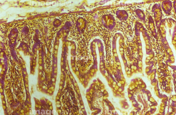
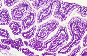
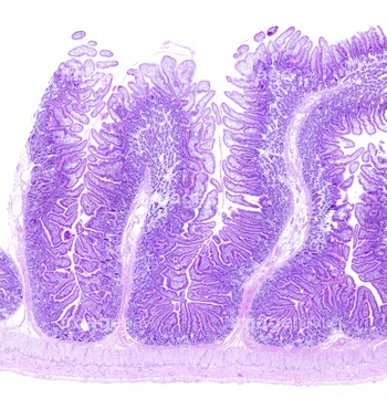
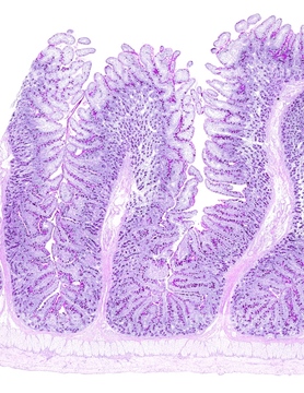
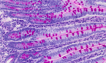
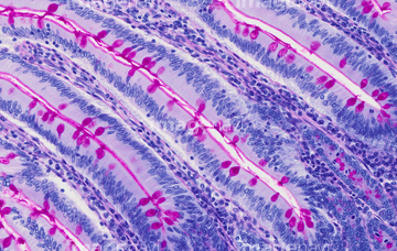
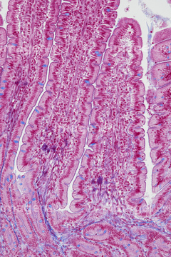
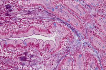
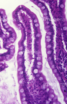
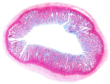
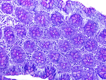
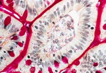
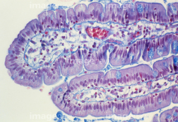
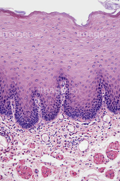
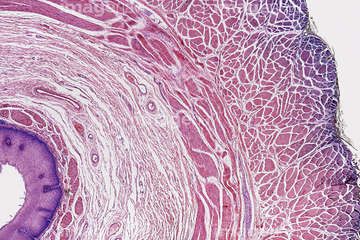
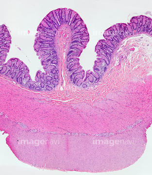
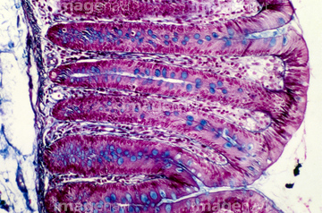
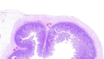
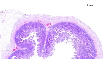
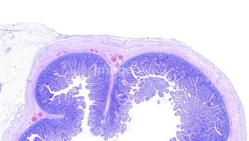
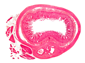
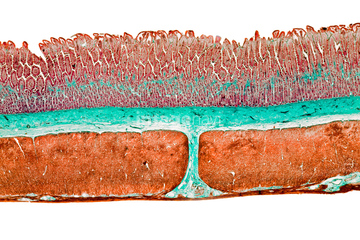
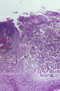
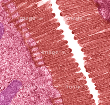
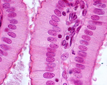
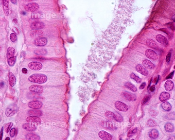
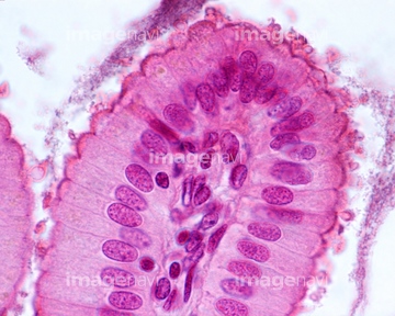
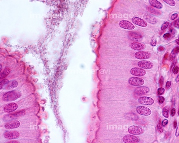
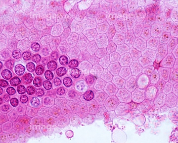
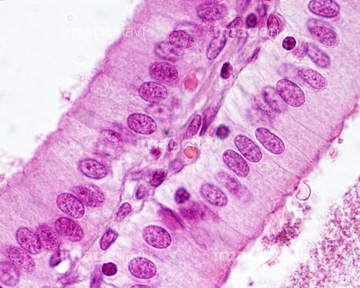
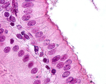
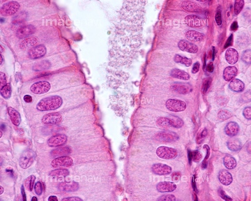
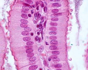
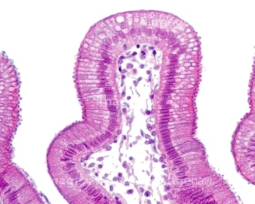
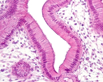
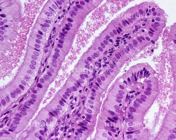
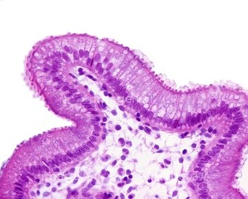
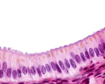
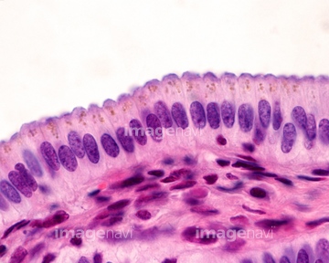
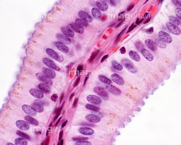
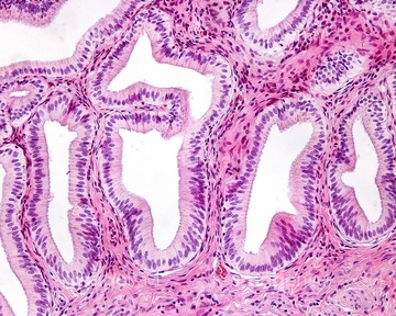
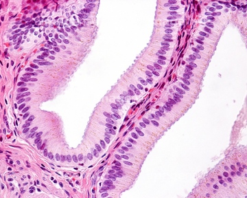
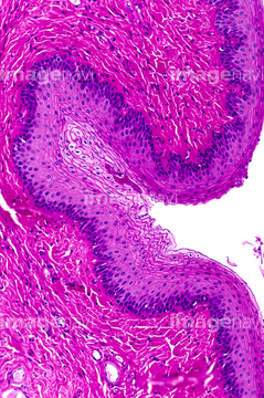
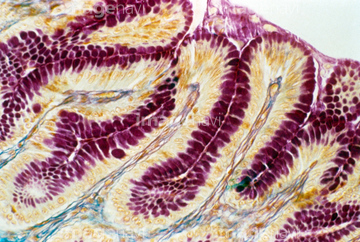
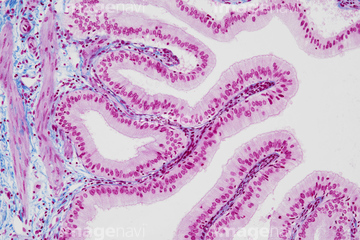
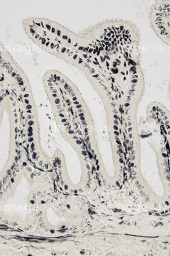
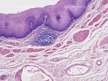
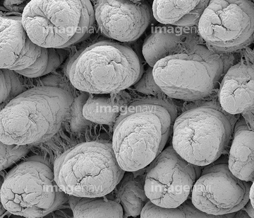
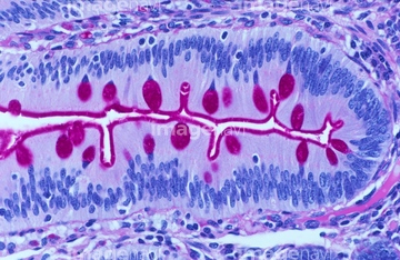
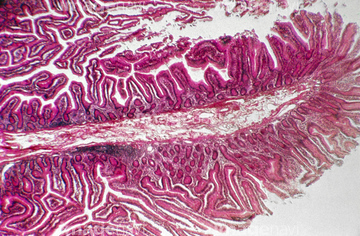
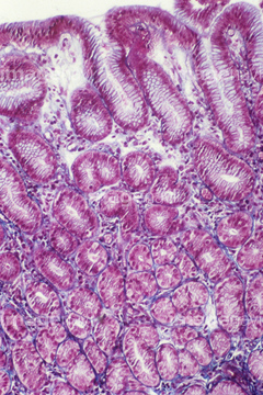
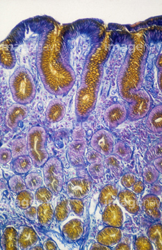
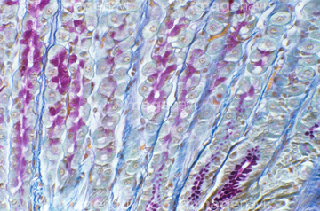
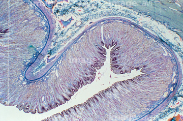
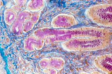
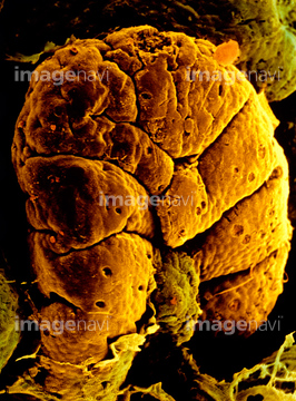
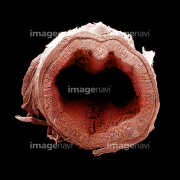
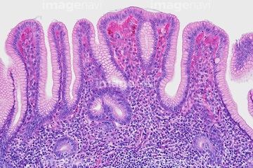
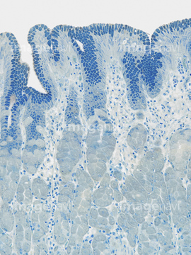
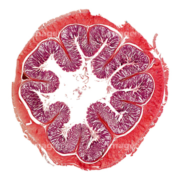
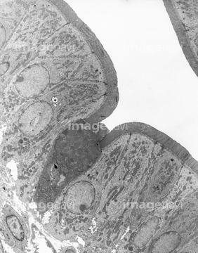
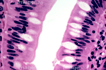
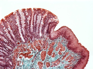
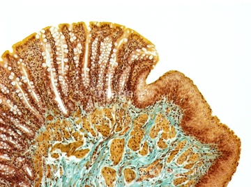
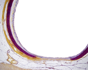
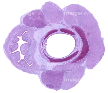
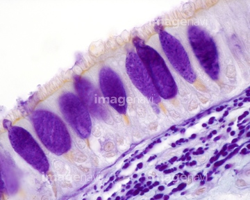
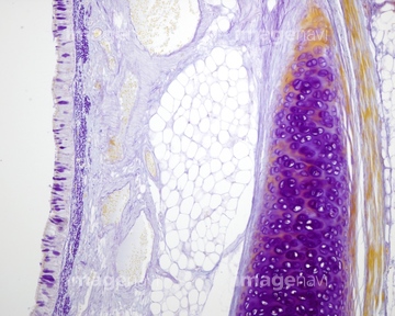
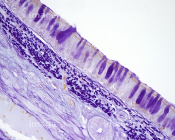
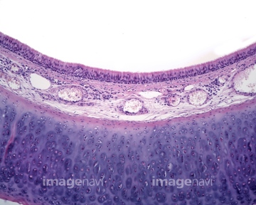
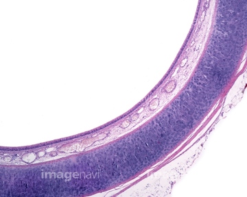
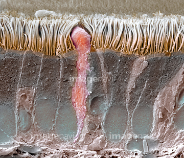
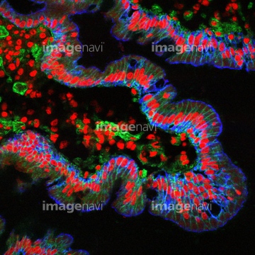
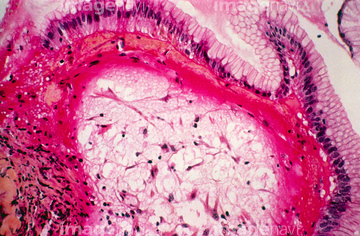
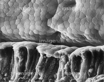
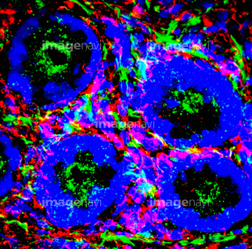
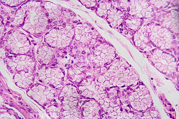
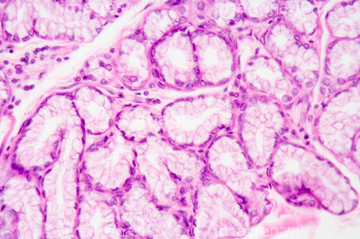
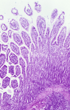
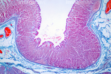
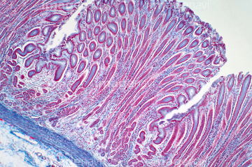
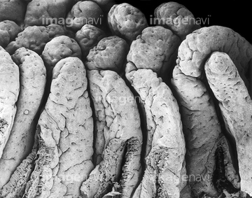
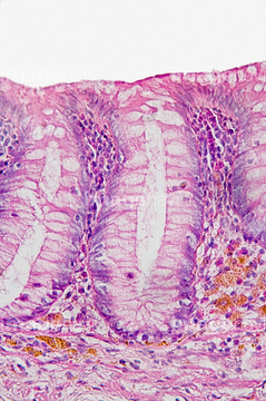
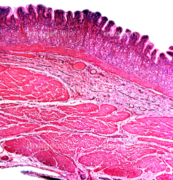
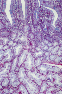
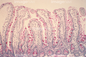
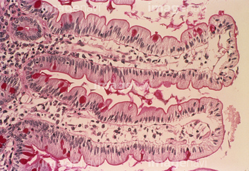
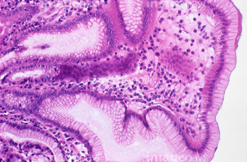
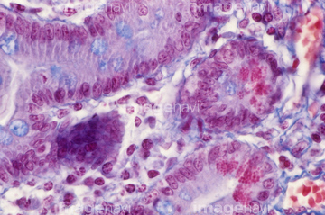
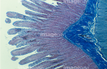
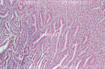
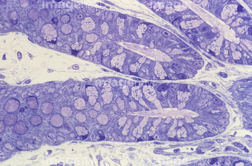
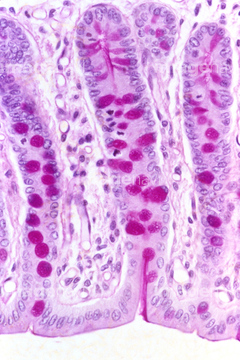
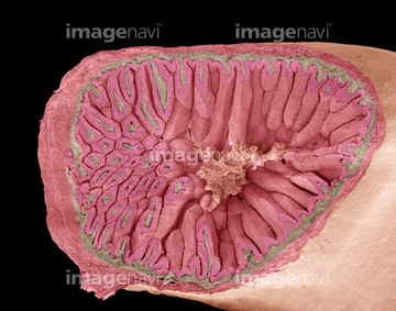
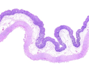
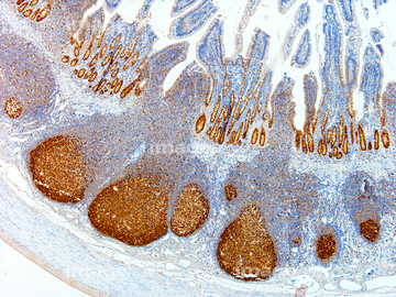
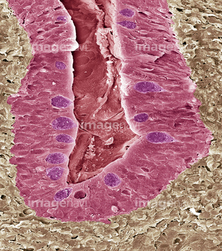
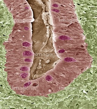
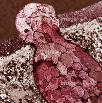
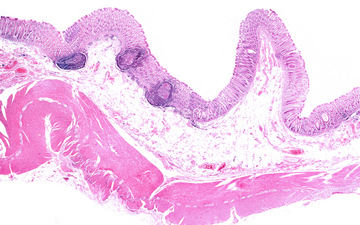
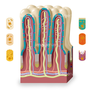
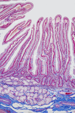
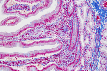
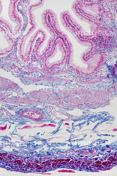
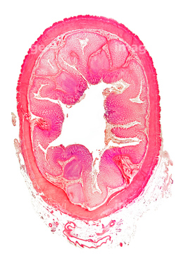
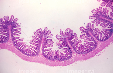
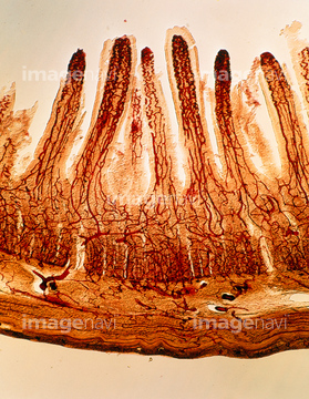
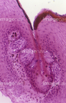
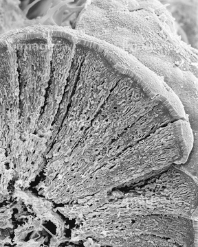
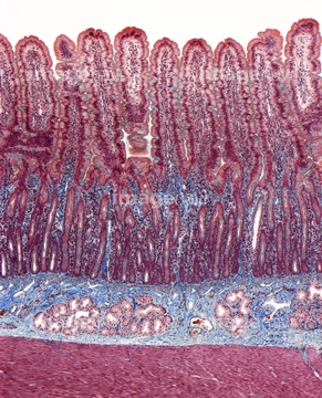
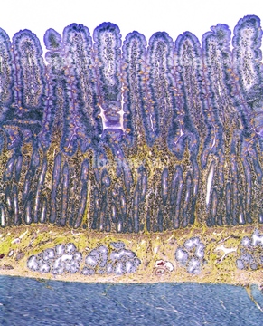
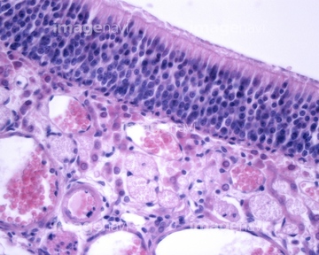
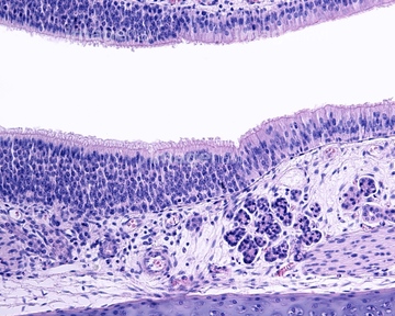
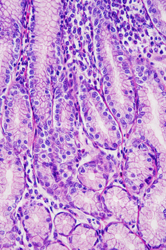
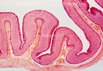
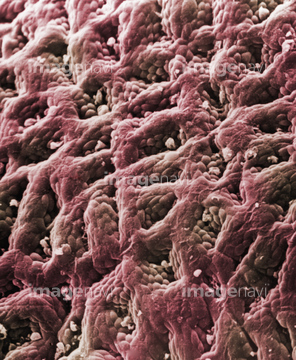
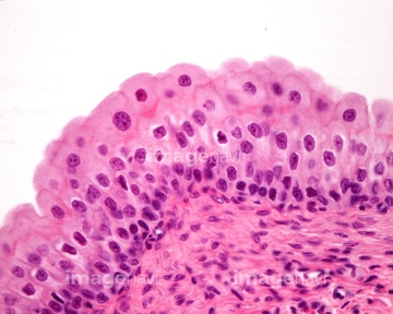
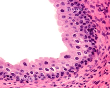
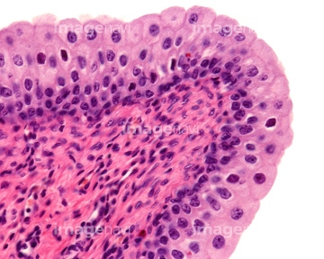
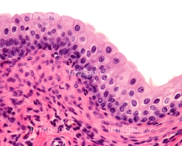
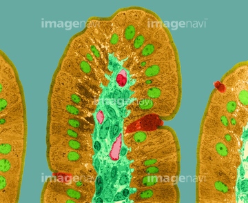
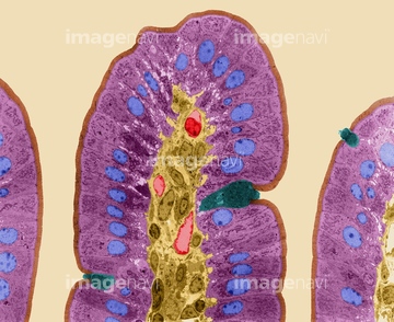
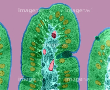
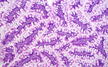
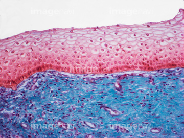
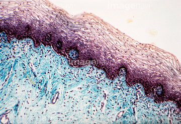
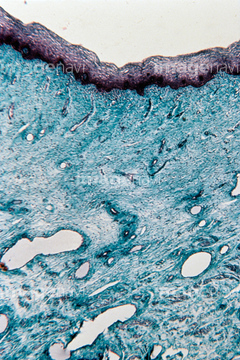
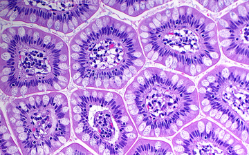
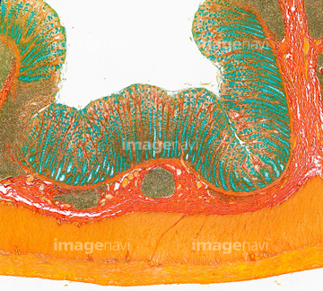
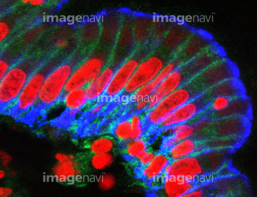
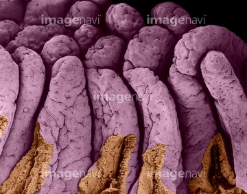
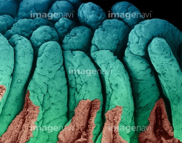
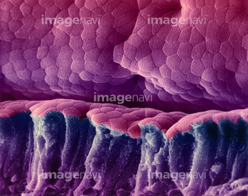
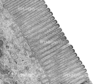
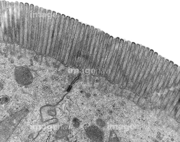
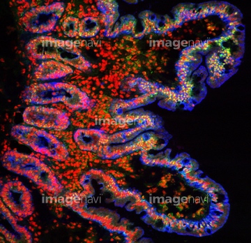
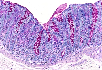
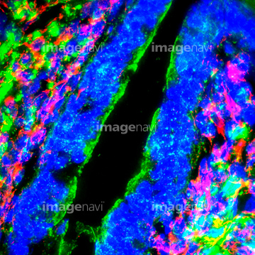
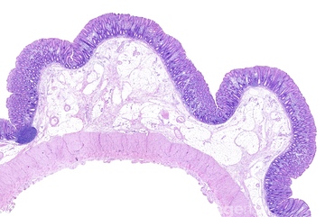
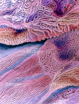
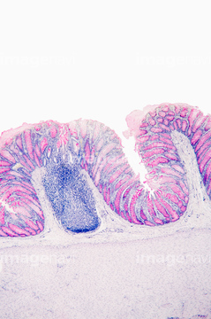
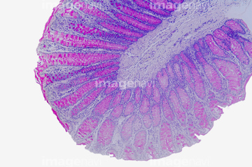
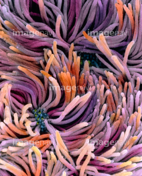
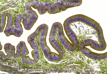
この検索結果には、Simple columnar epithelium, light micrograph、Gallbladder mucosa, light micrograph、Stratified squamous epithelium、Stomach lining, light micrograph、Gallbladder, light micrograph、Oesophagus, light micrographなどが含まれています。
64196734
64206819
64251229
64251226
64234476
64234477
64234478
64234479
64234667
64234718

64116645
64060933
64096008

64146135

64146275
64235009
64235010

64225083
64090172
64071989
64213913
64213914
64213915
17202471
17202472
64224938
64196715
64147742
64197478
64221011
64066084
64071988
64234509
64234510
64235008
64152547

64225140
64116647
64088314
64073272
64073273
64073274
64073275
64073276
64088319
64088323
64197489
64114561
64234280
64225131

64234840
64234841
64234843
64084027
64224843
64205896
64205907
64161677
64161678
64161681
64161683
64161684
64057593
64057594
64057595
64057596
64057597
64057598
64066076
64199159
64072028

64072029
64114553
64114554
64224937
20542530
20544099
64150338
64114552
64061536
64197486
64090152

64225133
64075661
64090479
64090480
64090490
64008744
64008745
64225094
64050457
64234473
64234474
64234508
64085267
64205888
64205892
64161259
64161268
64234842
64225092
64225256
17235956
64197456
64177447
64254349
64021554
64132258
64132996
64132997
64256459
64093993
64063011
64063015
64250037
64132948
64132949
64073343
64245044
64106063
64196735
64136447
64114573
64114574
64207219
| 次ページ |
