HOME > 写真 > 科学・テクノロジー > 科学 > DNA・細胞
10,000件の写真素材が検索されました。
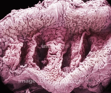
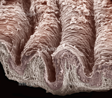
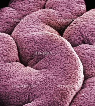
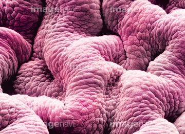
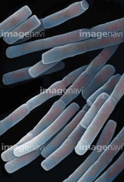
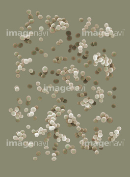
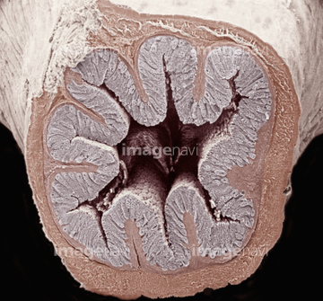
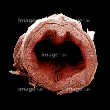
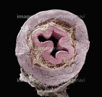
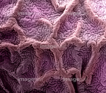
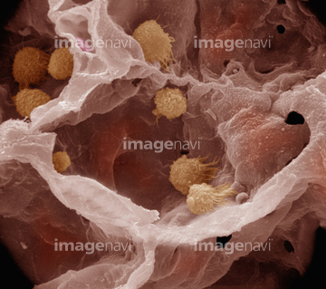
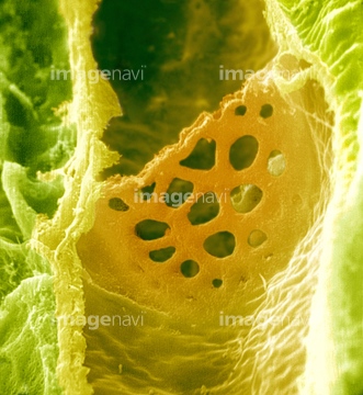
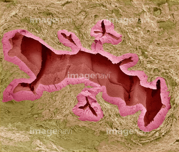
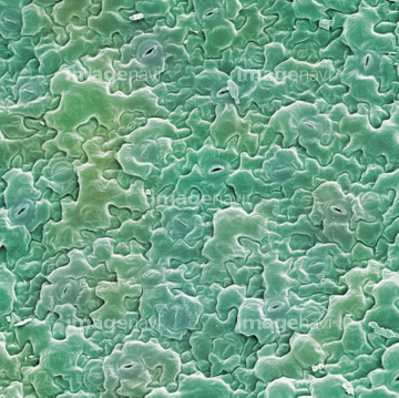
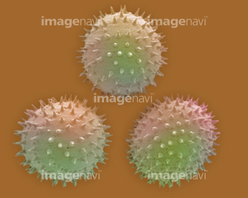
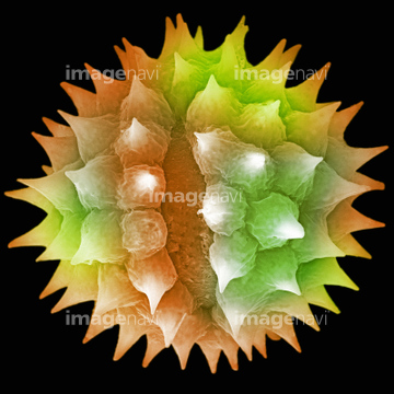
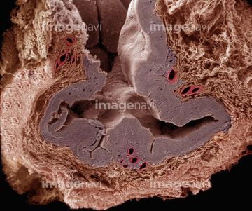
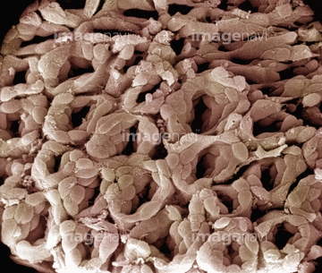
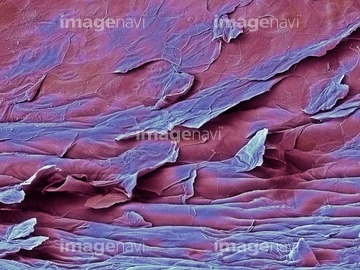
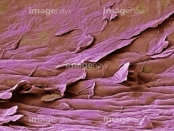
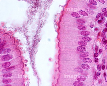
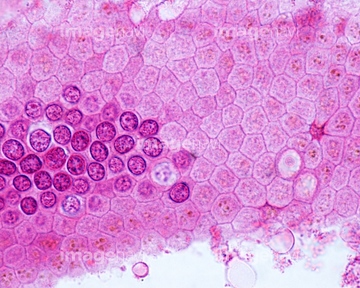
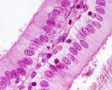
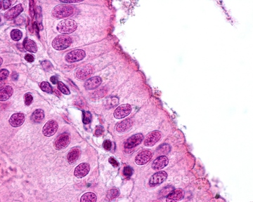
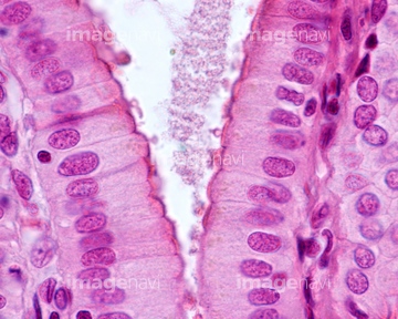
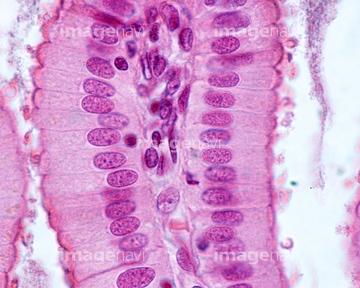
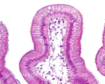
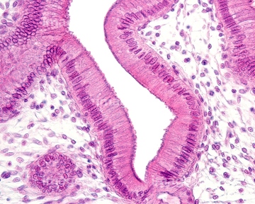
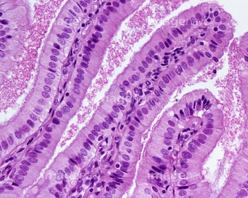
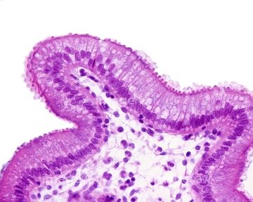
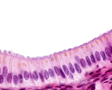
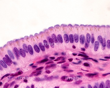
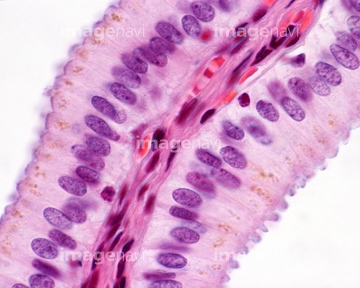
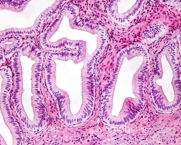
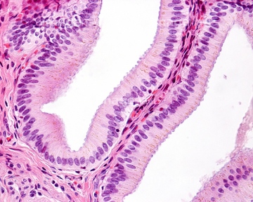
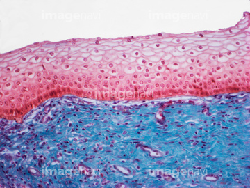
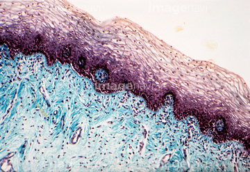
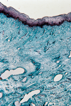
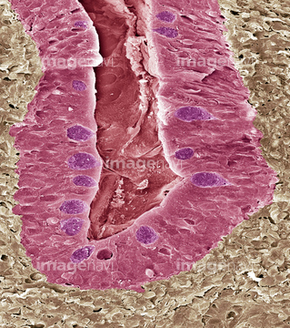
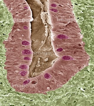
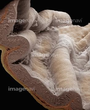
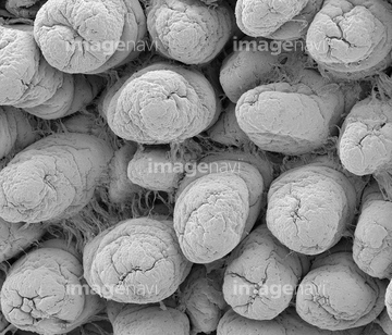
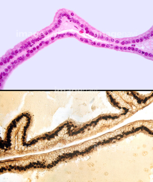
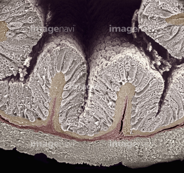
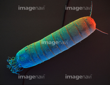
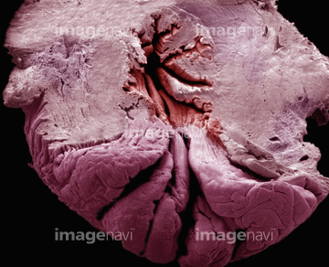
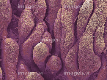
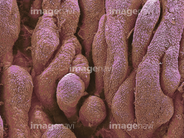
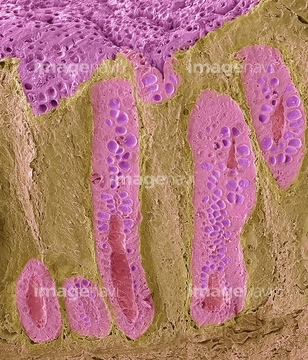
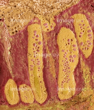
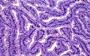
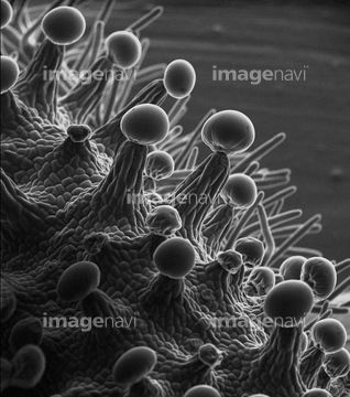
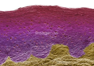
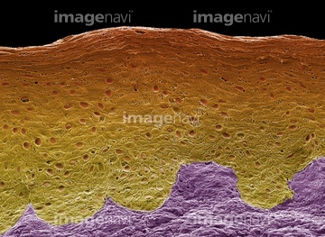
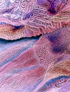
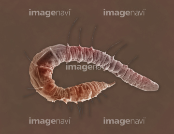
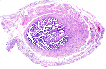
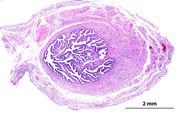
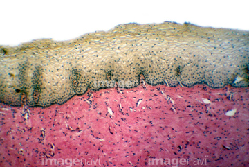
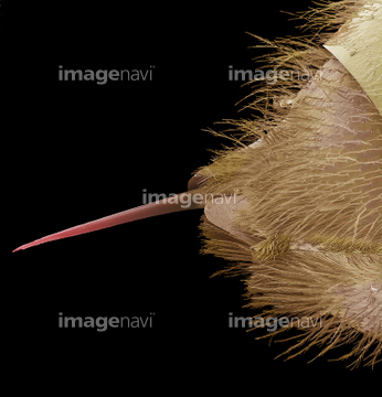
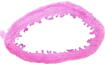
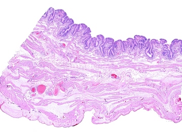
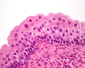
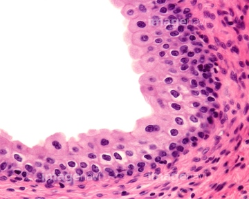
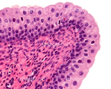
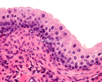
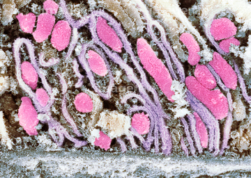
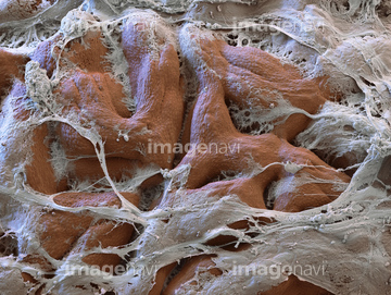
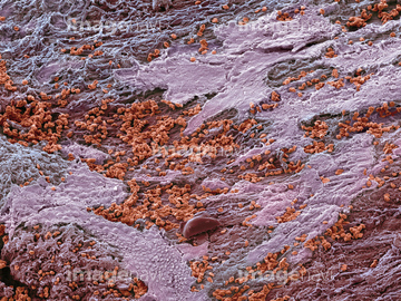
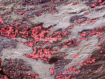
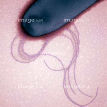
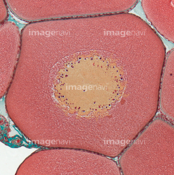
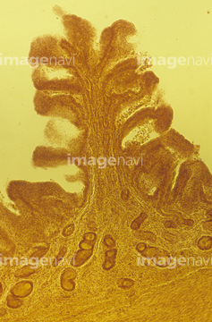
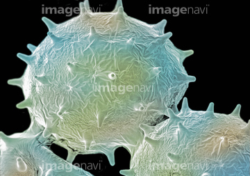
この検索結果には、Surface of pyloric stomach, mucous coat、Vaginal lining, SEM、Simple columnar epithelium, light micrograph、Gallbladder mucosa, light micrograph、Human vagina, light micrograph、Vaginal mucosa, light micrographなどが含まれています。
64225261
64225259
64225361
64225201
20548481
20548491
64235143
64235146
64235147
64235148
64115471
64115526
64115493
64114730
17246260
17246264
17246265
64115520
64115594

64225243

64146135

64146275
64224880
64225175
64225131
64225192
64225260
64225190
64225244
64061548
64115490
64115595
64115597

64115600

64225202
64190237
64215656
64221011
64093993
64063011
64063015
64008744
64008745
64225256
64224854
64111600
64111608
64111633
64077392
64061547
64030477
64030789
64225091
64225242
64225257
64115535
64115536
64115521
64115593
64225176
64152547

64225140
64029976
64259017
64259018
64195869
64195893
64245072
20507330
20507334
64136447
64225101
64256993
64065178
64021756
64065986
64250182
64250202
64114169

64225083
64225088
64225129
64225130
64225132

64225133
64225151
64225152
64197456
64037351
64176856
64176857
64176858
64225092
64008746
64008751
64225086
64225264
18597771
64245075
17202471
17202472
64115596
17281852
17281881
17281895
64225265
64235009
64235010
64205395
64247719
64260716
64260718
64261830
64261831
64261837
64115602
64088773
64088787
64224945
64114732
64114733
64224879
64077495
64076289
64076290
64183414
64008178
64013157
64013158
64031148
64256051
64256052
64256053
17234583
64111617
64111643
64152612
| 次ページ |
