HOME > 写真 > 医療・福祉 > 医療 > 病院・クリニック
10,000件の写真素材が検索されました。
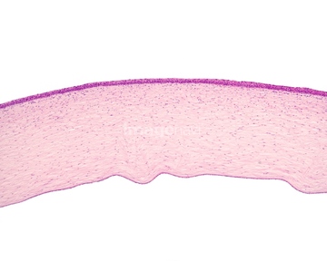
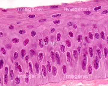
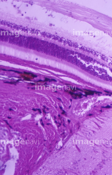
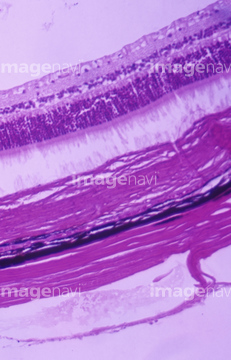
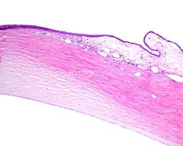
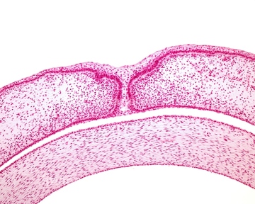
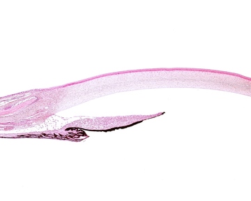
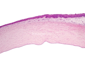
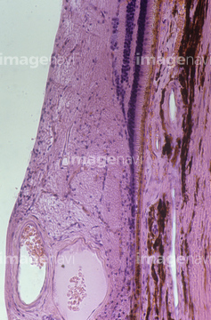
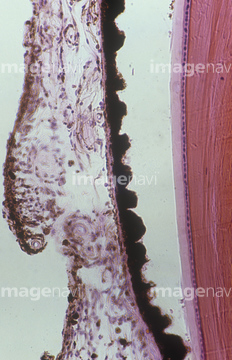
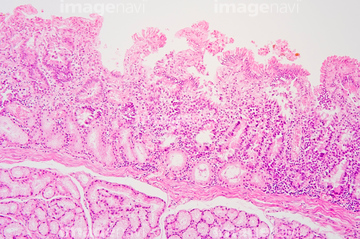
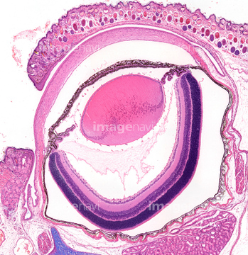
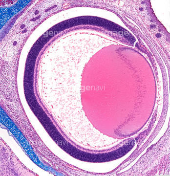
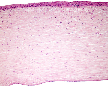
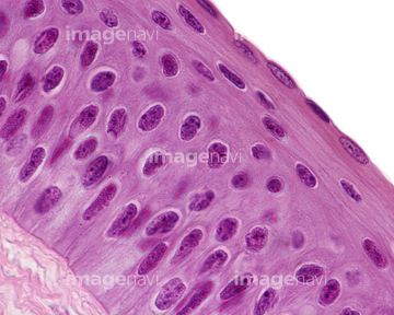
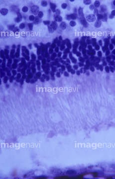
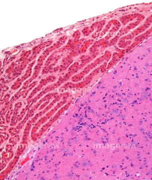
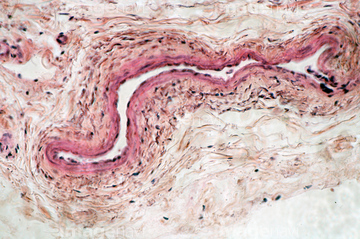
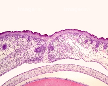
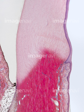
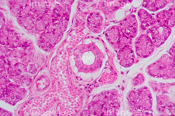
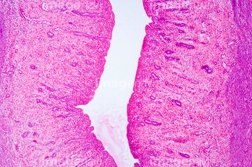
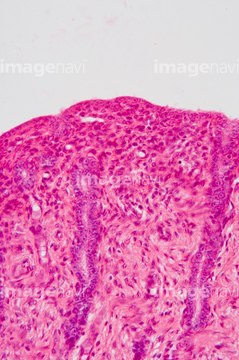
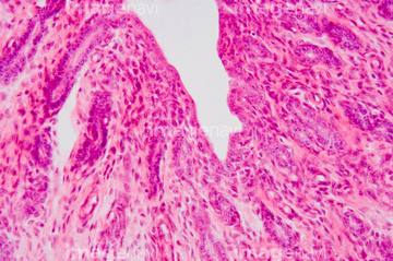
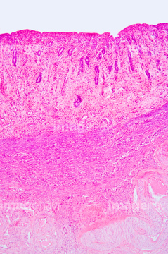
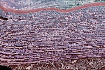
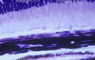
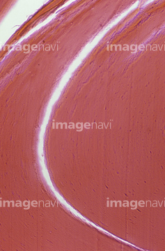
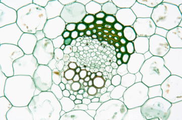
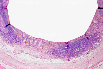
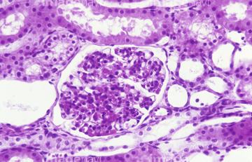
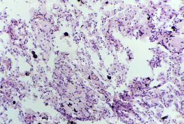
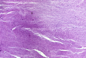
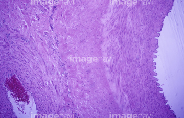
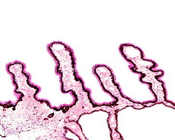
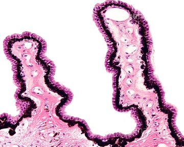
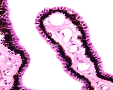
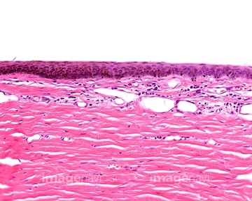
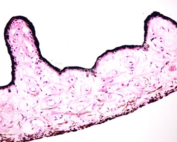
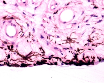
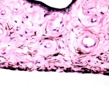
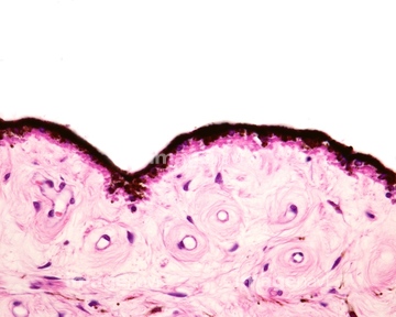
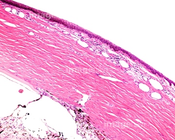
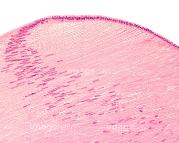
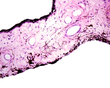
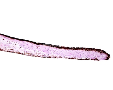
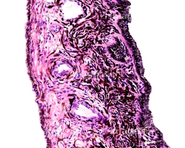
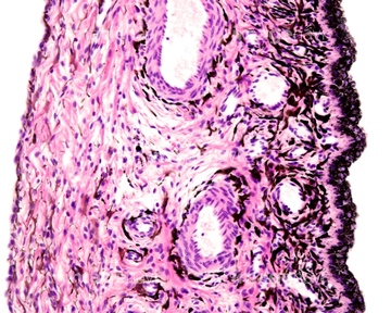
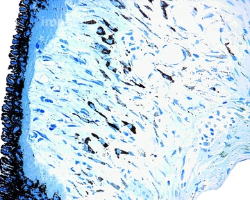
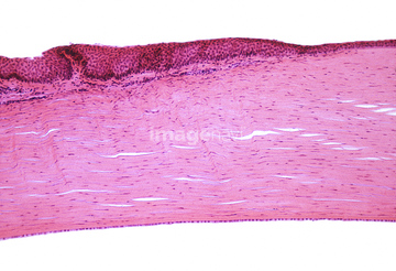
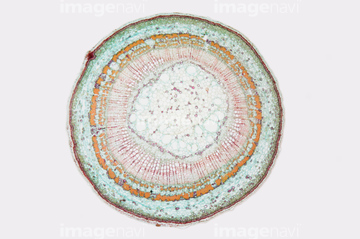
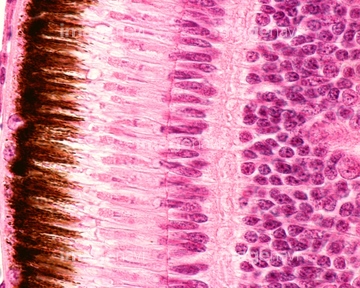
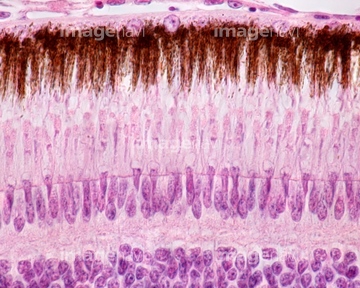
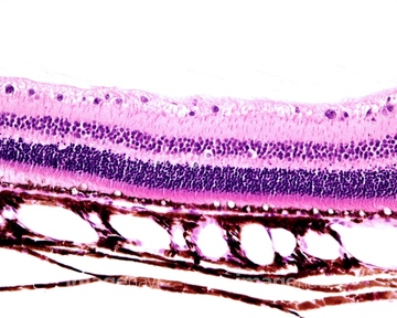
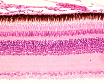
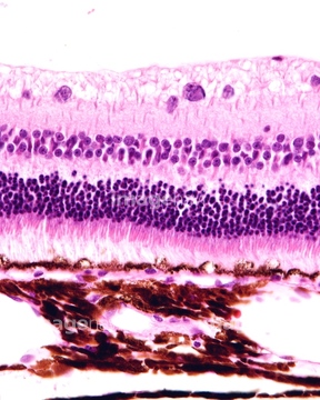
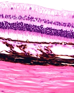
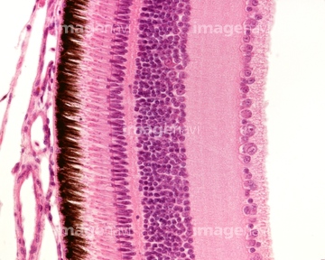
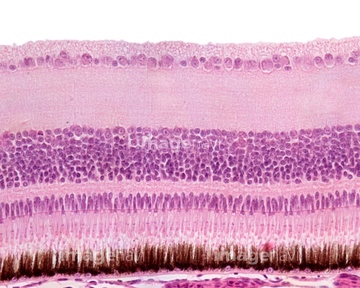
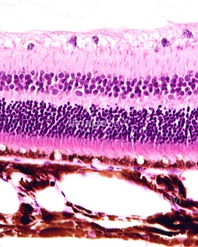
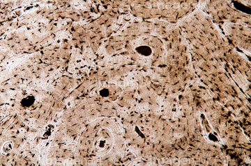
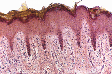
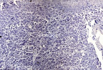
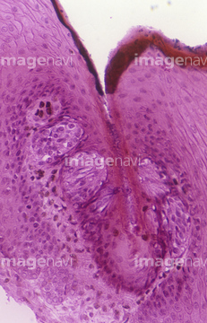
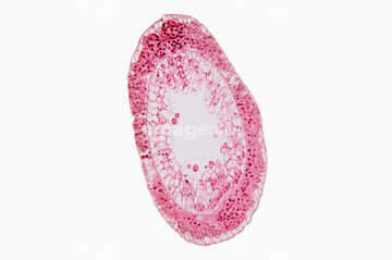
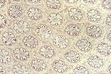
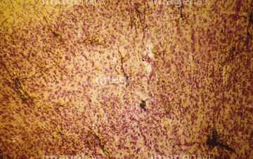
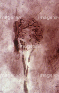
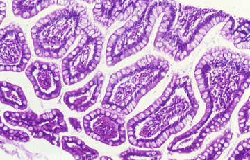
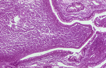
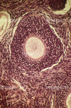
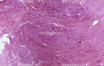
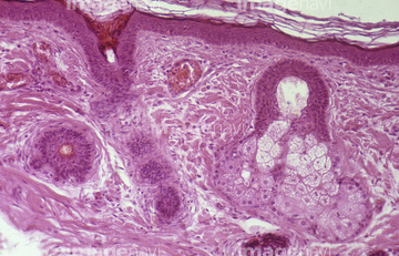
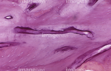
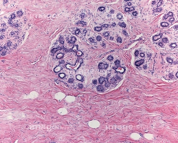
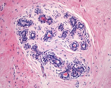
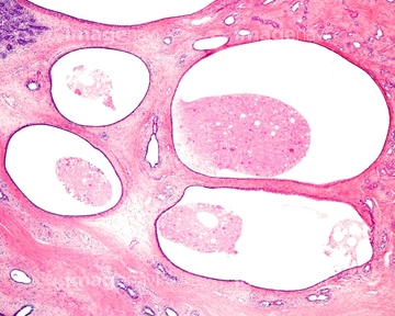
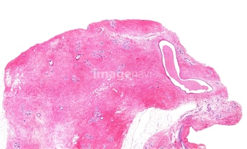
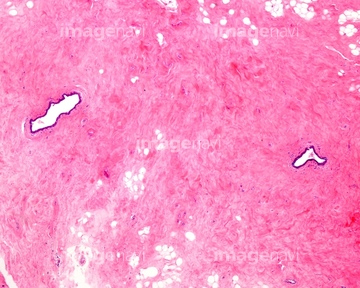
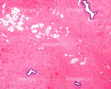
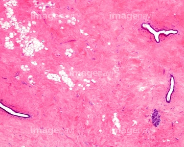
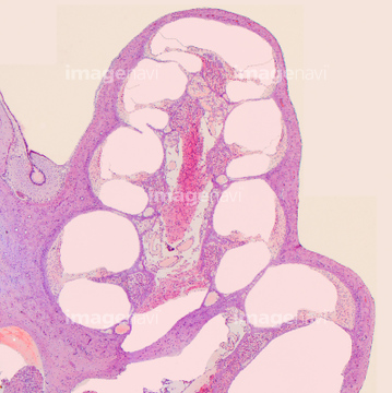
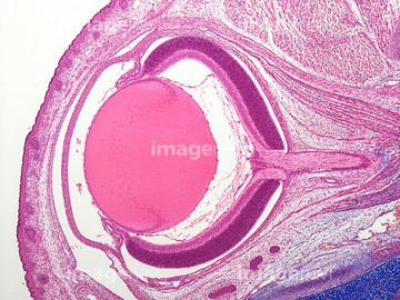
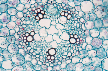
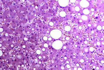
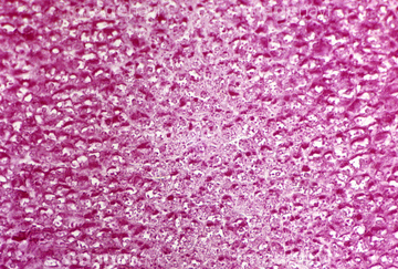
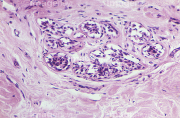
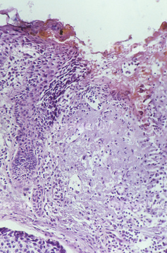
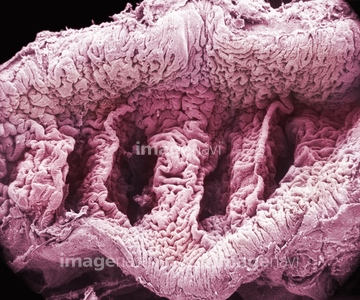
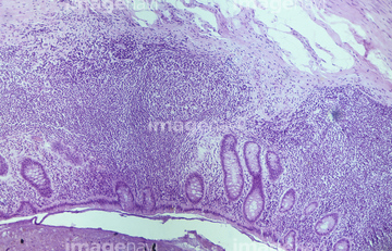
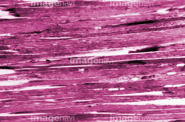
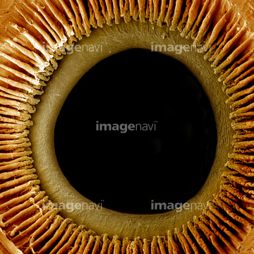
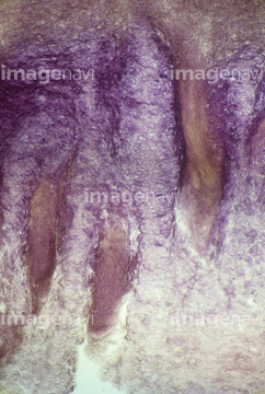
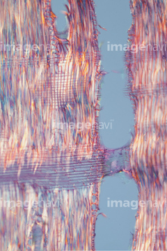
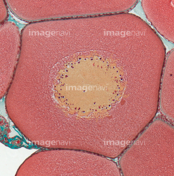
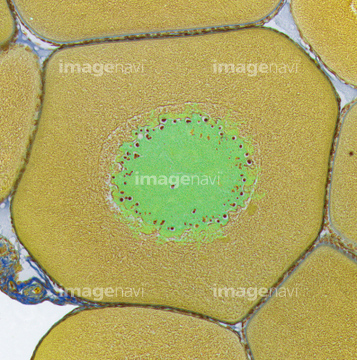
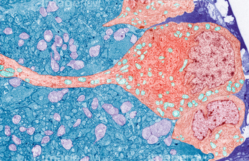
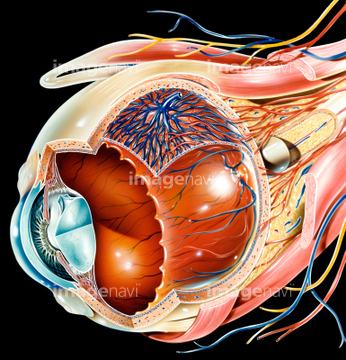
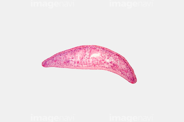
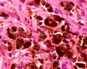
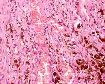
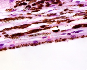
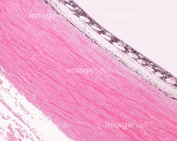
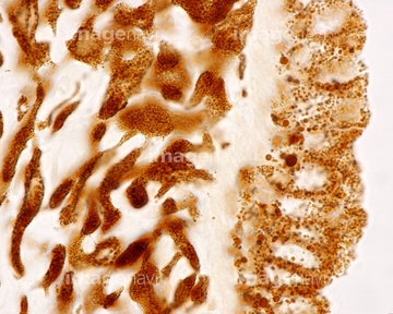
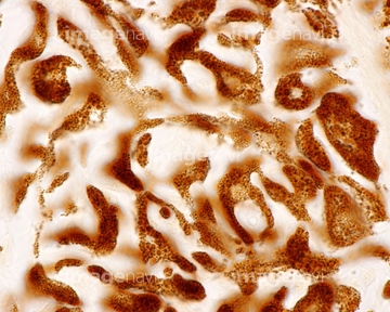
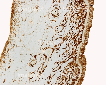
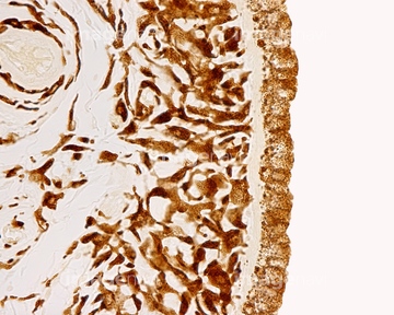
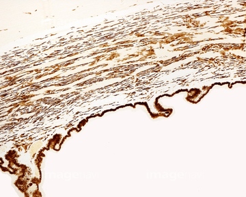
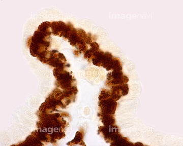
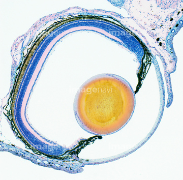
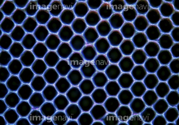
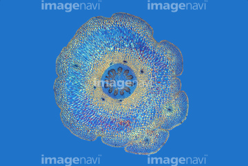
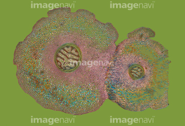
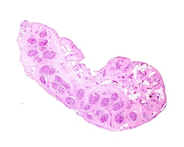
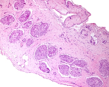
この検索結果には、Cross-section of the human vagina. LM、Cross-section of human vagina. LM、Cornea, SEM、Section of the eye retina pigment layer、Section of the eye lens LM X80、Cross-section of a plant vascular bundleなどが含まれています。
64114524
64114522
64114523
64190709
64190710
64224839
64198955
64198956
64198958
64198964
64190713
64225213
64114549
64090168
64090170
64260240
64260244
64225221
64114570
64114571

64114521

64148801
64148804
64115571
64198945
64234648
64114552
64114553
64114554

64114540
64114526
64114529
64039302
64225212
64114551
64115581
64114573
64114574
64114561
64224953
64224954
64224932
64225232
64225233
64190681
64190682
64190689
64190690
64190691
64190692
64190693
64190696
64190712
64194306
64216423
64216439
64216440
64213935
64190721
64190723
64190724
64190726
64114577

64146135

64146275
64055576
64224938
64224945
64224950
64224951
17235966
64180281

64213859
64213860
64213861
64088778
64073358
64224907
64224928
64225231
64225261
64224942
64225132
64225203
64088773
64088787
64056270
64067989
64114566
64198959
64198963
64198966
64198967
64198968
64198969
64198973
64216434
64216435
64225224
64090169
64096078
64131225
64190680
64197461
64205318
64207667
64213894
| 次ページ |
