HOME > 写真 > 科学・テクノロジー > 科学 > DNA・細胞
10,000件の写真素材が検索されました。
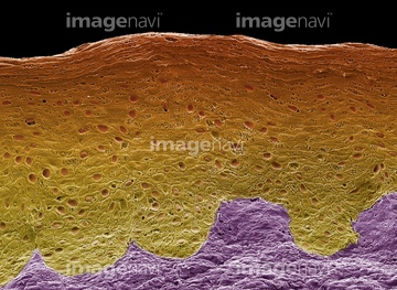
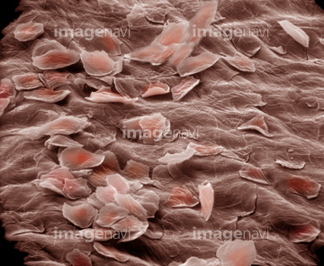
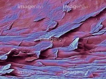
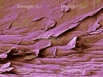
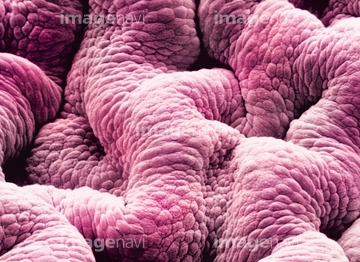
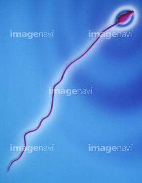
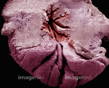
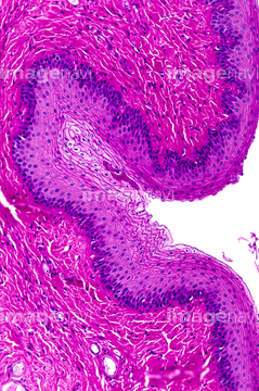
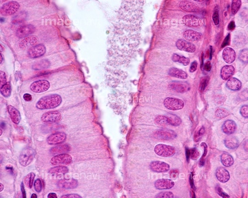
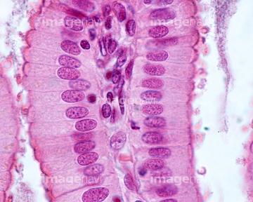
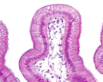
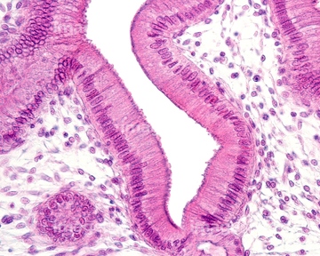
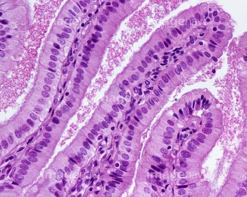
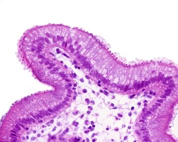
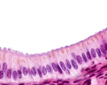
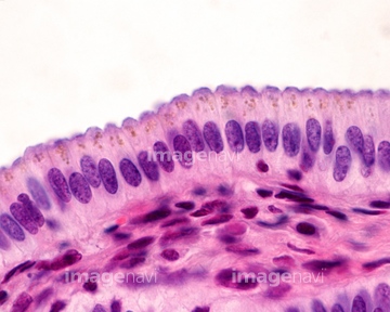
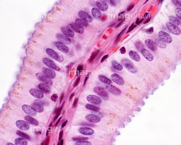
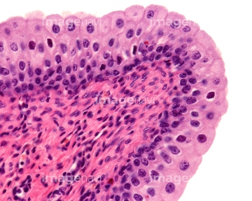
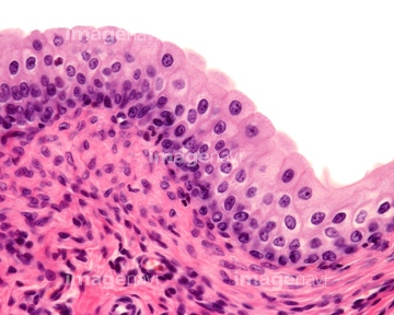
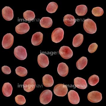
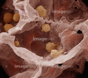
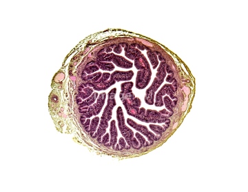
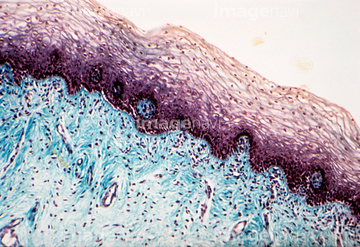
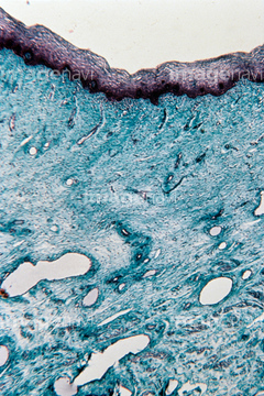
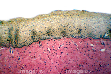
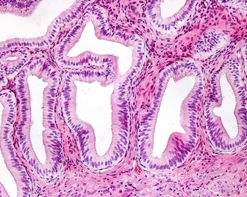
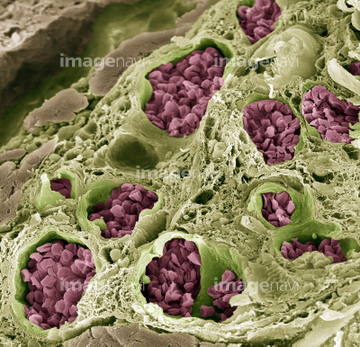
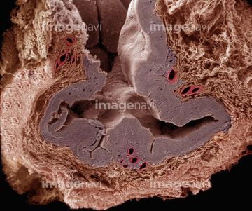
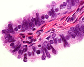
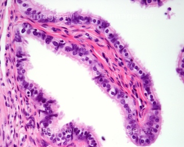
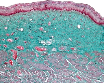
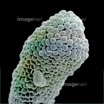
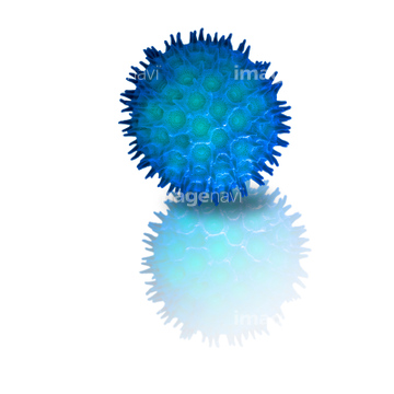
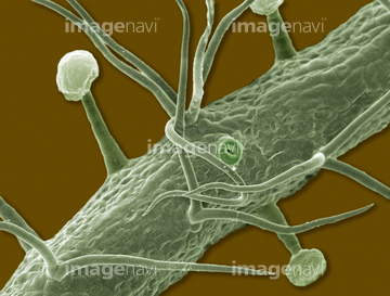
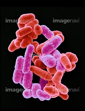
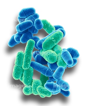
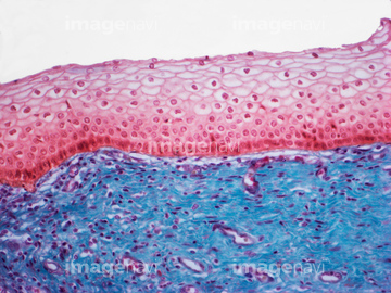
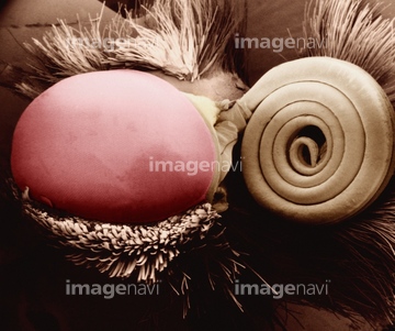
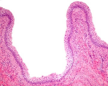
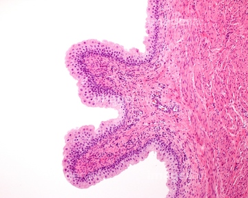
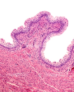
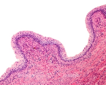
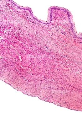
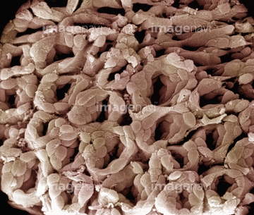
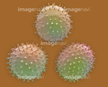
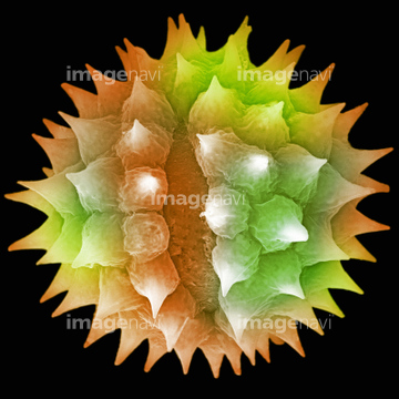
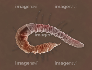
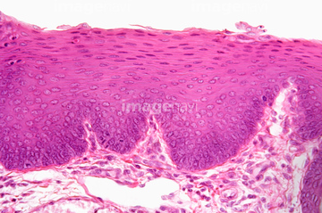
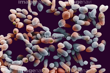
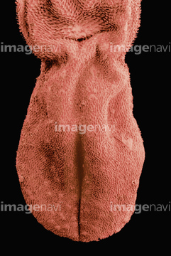
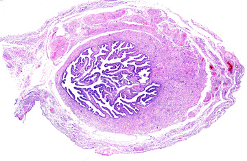
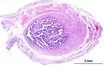
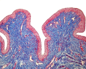
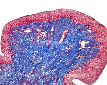
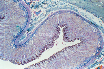
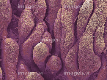
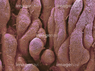
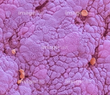
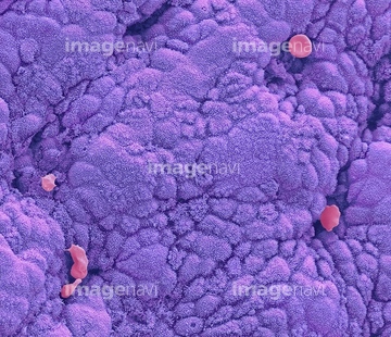
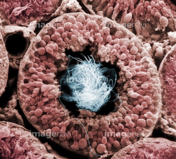
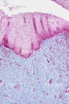
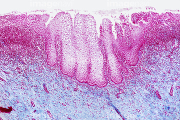
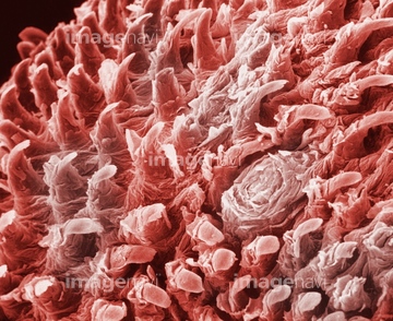
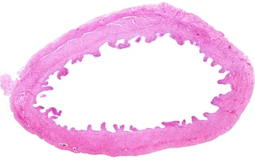
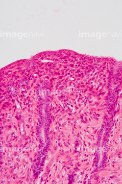
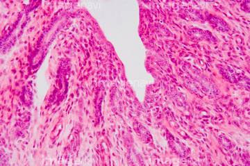
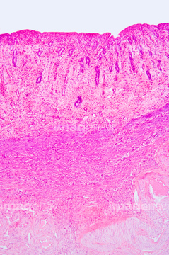
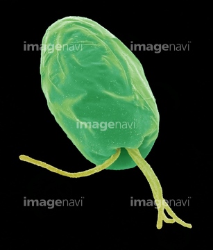
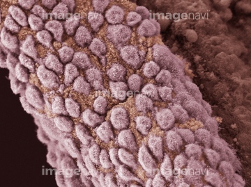
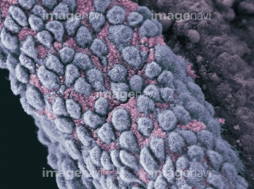
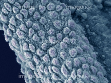
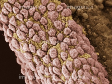
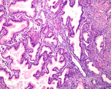
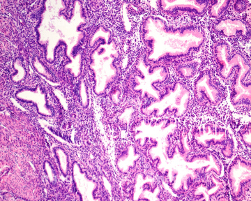
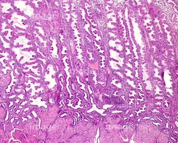
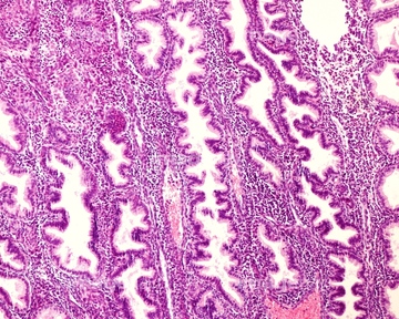
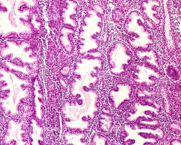
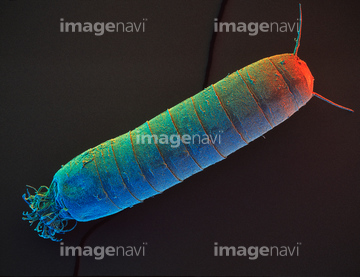
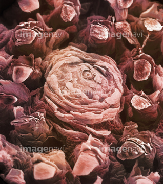
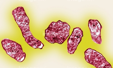
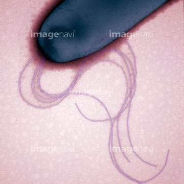
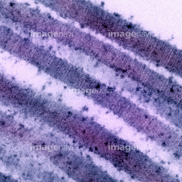
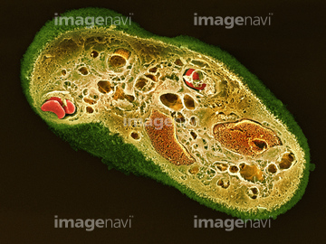
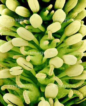
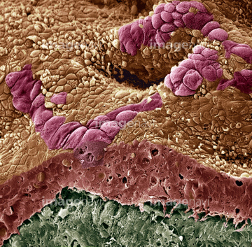
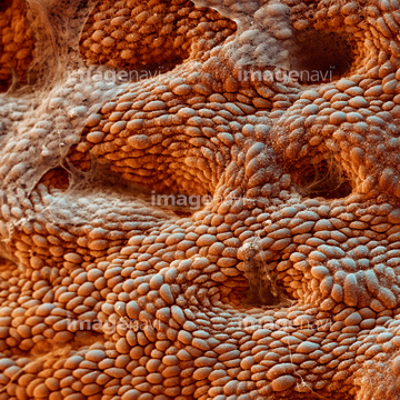
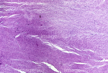
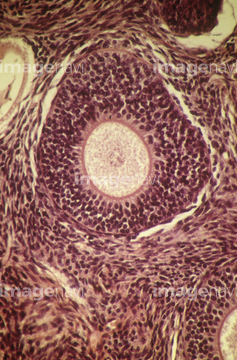
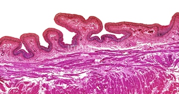
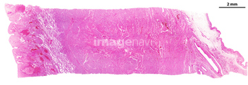
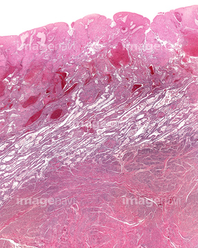
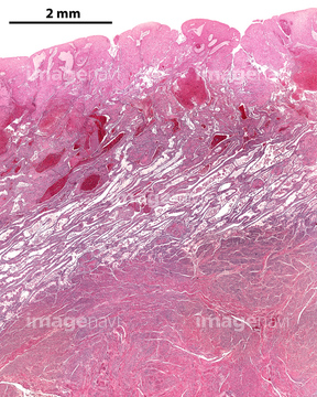
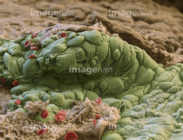
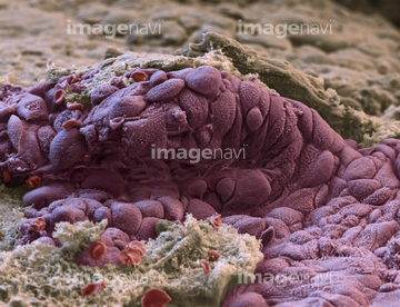
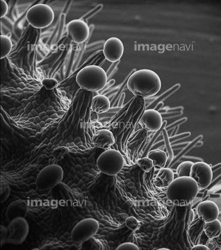
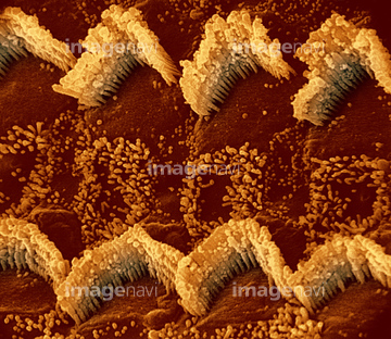
この検索結果には、Vaginal wall, light micrograph、Stigma of Penta lanceolata. SEM、Morning Glory pollen grain、Black Walnut (Juglans nigra)、Pine Pollen. SEM、(Bacillus megaterium) Bacteriaなどが含まれています。
64225261
20507330
20507334
64225201
64190237
64215656
64225259
64225361
64066084
64115471
64225190
64063011
64063015
64065986

64146135

64146275
64221011
64008744
64008745
64008746
64008751
64225202
64177447
64114730
64115520
64115526
64115594
64224880
64225175
64115493
64225260
64225131
64225192
64225189
17246260
17246264
17246265

64093993
64225243
64225244

64196734
64115490
64115595
64225101
64115600

64225140
64114561
64224854
64088791
64093995
64206819
64073272
64073273
64073274
64073275
64073276
64084027
64259017
64259018
64195821
64195822
64195869
64195893
64056751
64235141
64235142
64225257
64151681
64151682
64225086
64225091
64225242
64225264
64225151
64197456
64114169
64176856
64176857
64176858
64225092

64114526
64114529
64124825
64124832
64124833
64124844
64124852
64111590
64111635
64116370
64160041
64160042
64160045
64129016
64077491
64076340
64076341
64256051
64256052
64256053
64225183
64190619
64190620
64190621
64190622
64029976
64225082
64115602
64115597
64076566
64006251
64006281
64006282
64006283
64006286
64008081
64056745
64224945
64152547
64225256
17202471
17202472
64207217
64207264
64176910
64176911
64224897
64245072
64225069
| 次ページ |
