HOME > 写真 > 科学・テクノロジー > 科学 > DNA・細胞
10,000件の写真素材が検索されました。
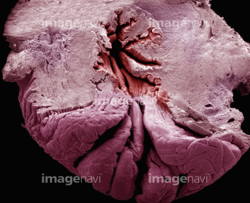
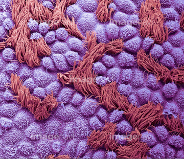
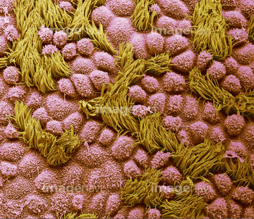
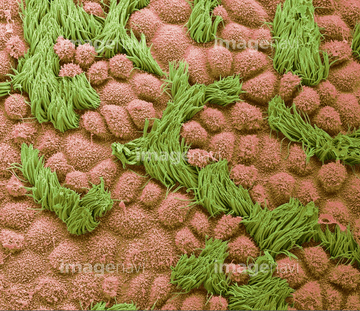
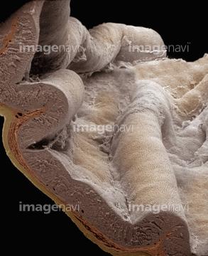
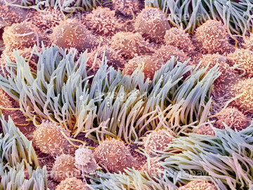
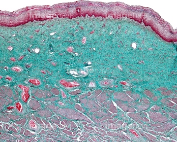
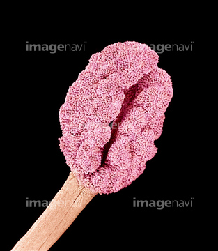
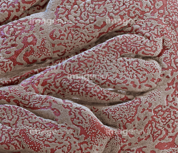
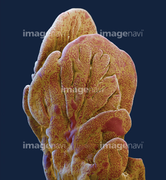
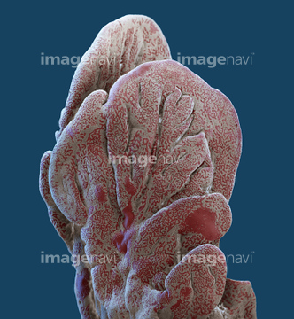
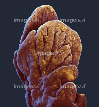
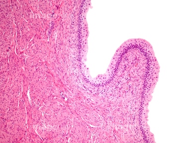
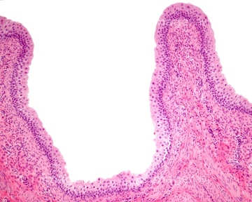
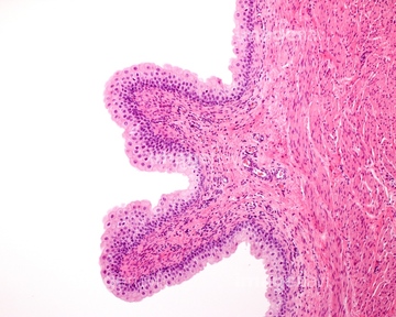
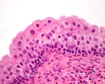
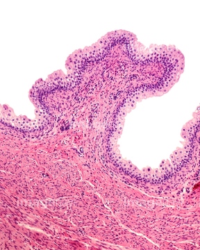
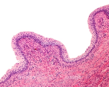
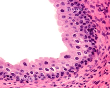
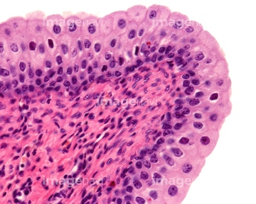
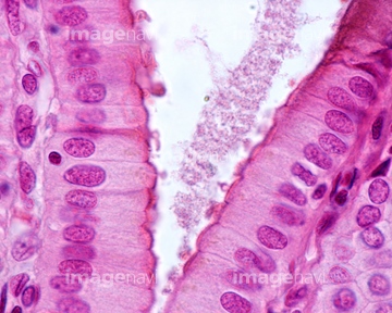
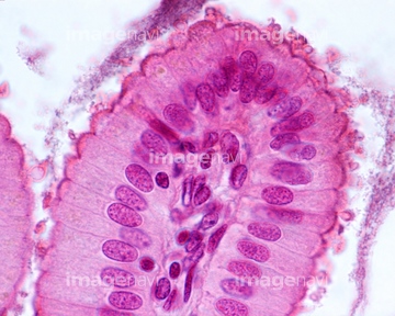
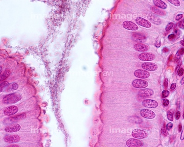
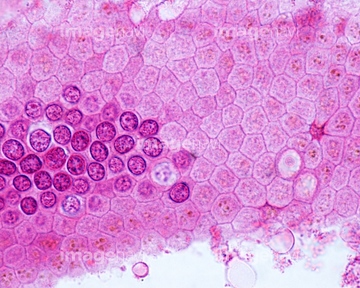
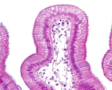
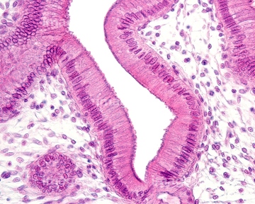
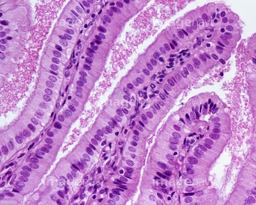
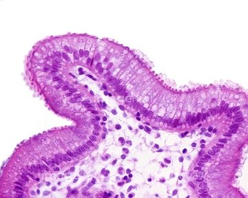
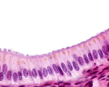
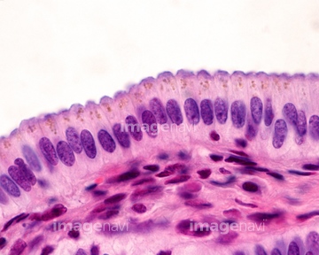
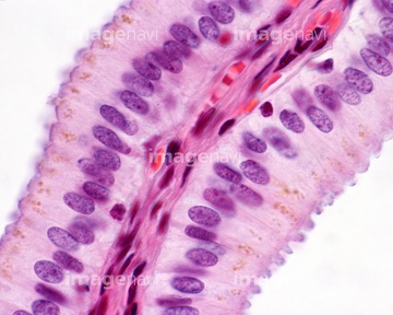
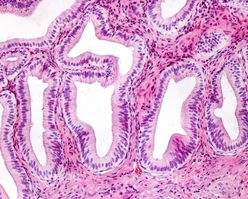
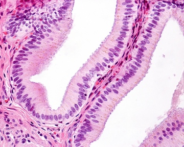
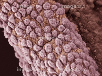
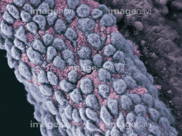
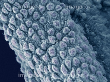
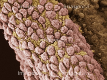
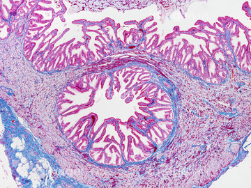
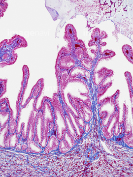
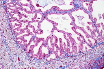
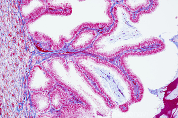
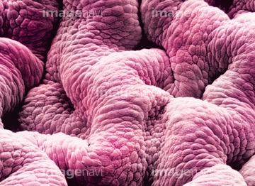
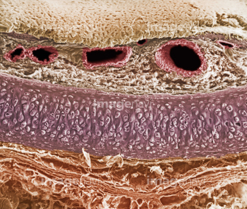
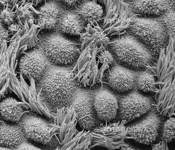
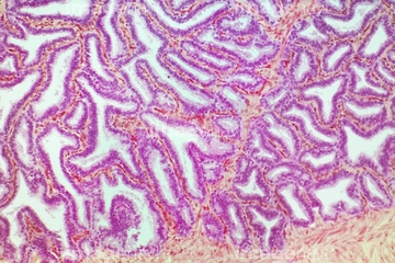
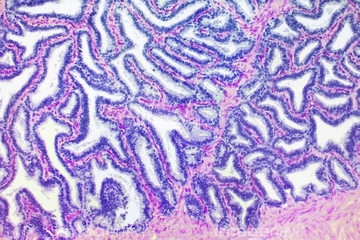
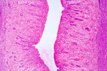
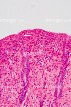
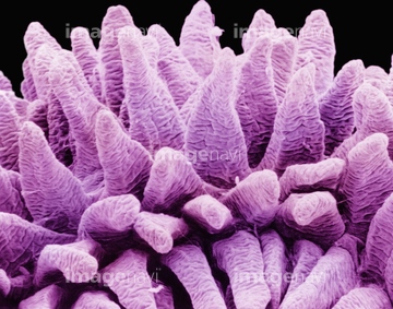
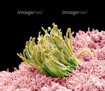
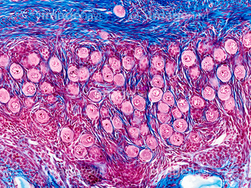
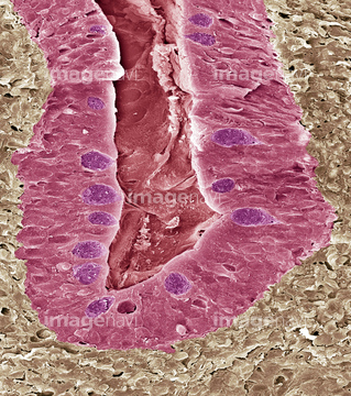
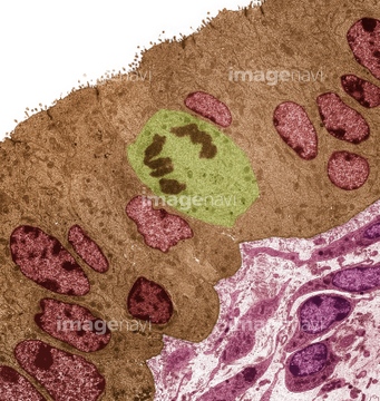
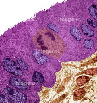
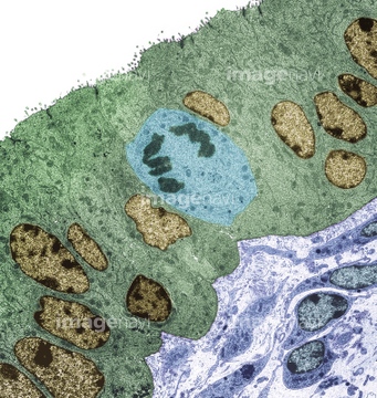
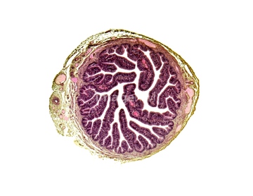
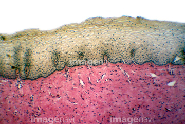
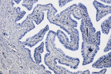
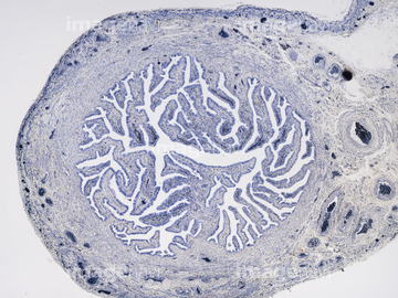
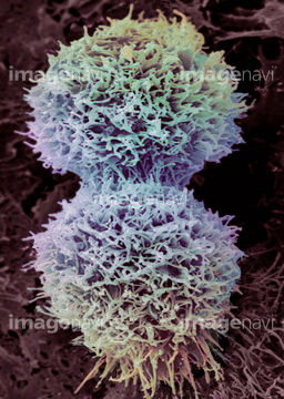
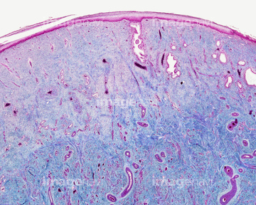
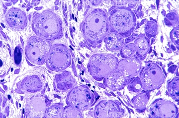
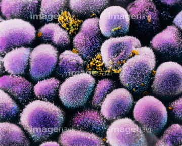
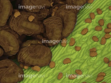
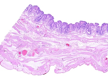
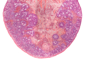
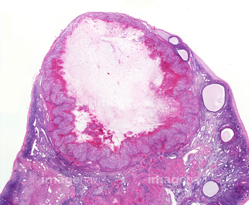
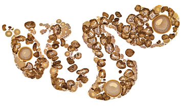
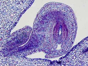
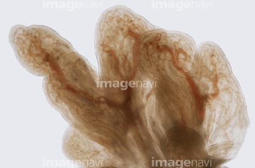
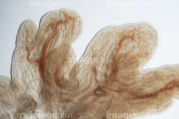
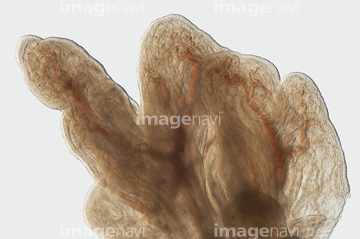
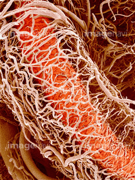
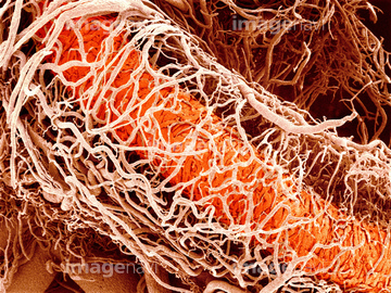
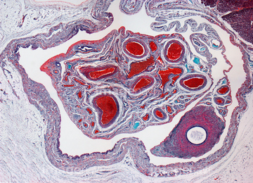
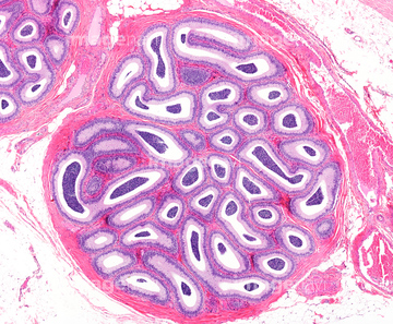
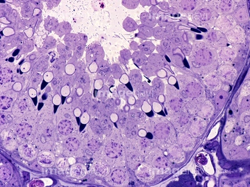
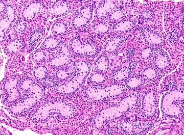
この検索結果には、Simple columnar epithelium, light micrograph、Gallbladder mucosa, light micrograph、Uterus lining, SEM、Villi in mammal small intestine、Seminal vesicle, light micrograph、The mucosa of the urinary bladderなどが含まれています。
17290287
17290293
17290301
64225202
64190237
64056751
64177447
64082467
64077491
64076340
64076341
64199723
64199724
64199737
64199738
64199739
64199740
64199741
64199742
64199743

64146135

64146275
64221011
64190619
64190620
64190621
64190622

64225140
64235143
64235146
64235147
64235148
64225259
64225189
64152541
20507330
20507334
64215656
64225199
64225131
64192873
64192878
64111600
64111608
64111633
64077495
64076289
64076290
20548481
20548491
64114526
64114529
64093975
64093976
64225256
64075100
64060928
64008744
64008745
64008746
64008751
64097155
64203396
64203413
64203445
64084027
64196734
64206819
64066084
64030738
64256043
64256044
64256045
64092932
64154365
64155028
64157156
64157166
64106441
64106443
64106447
64106452
64225261
64063011
64063015
64065986
64234481
64234484
17201282
64192859
64192866
64192886
64225260
64234961
64116657
64060681
64148622
64209491
64209492
64209493
64114169

64225083

64225133
64225151
64225152
64225200
64197456

64088772
64098225
64085234
64263386
20523344
20523360
64198564
64260716
64260718
64261830
64261831
64261837
64265112
64265117
64265122
64265150
64265153
64265169
64225071
64090434
64092136
64106444
64011864
64225158
64225201
64093999
64098234
64106072
| 次ページ |
