HOME > 写真 > 科学・テクノロジー > 科学 > DNA・細胞
10,000件の写真素材が検索されました。
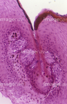
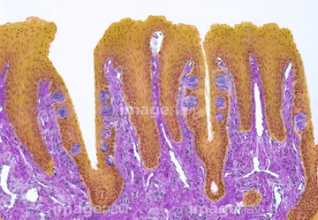
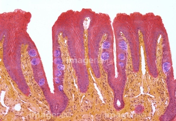
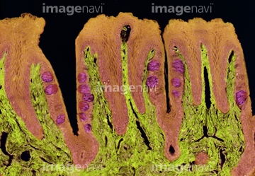
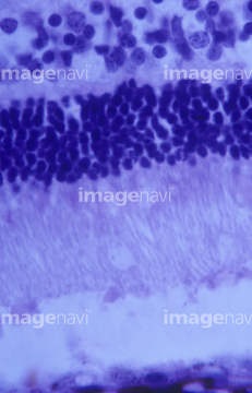
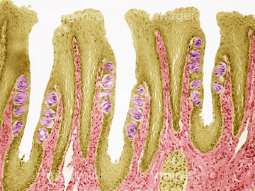
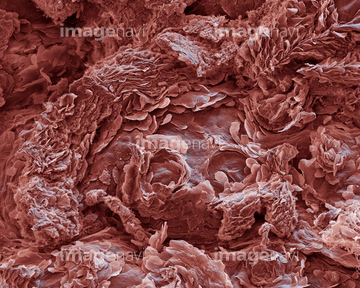
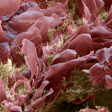
この検索結果には、Tongue surface, SEM、Tongue papillae, SEM、Section of the retina fovea centralis. LM、Section of the central optic disc. LM、Cat tongue, polarised light micrograph、Section of the optic disc of the eye. LMなどが含まれています。
20532887
20532889
20532894
64068329
64213898
64213899
64213900
64213901
64213902
64235184
64235186

64085273
64065982

64225232
64225233
64235183
64082525
64076315

64076316
64076317
64114561
64114526
64114529
64092969
64059771
64010899

64010900
64010901
64010902
64010903
64010904
64010905
64011856
64011858
64011860
64011861
64011862

64146135

64146275
64114552
64114573
64114574
64224942
64225221
64008177
64074874
64047103
64225213
64003130

64003131
64225212
64224907
64224932
64224938
64225231
64224953

64224841
64115571
64224904
64224954
64035025
64035536
64035537
64114551
64115581
64225203
64190691
64213935
64047104
64114540
64114570
64114571
64114577
64167101
64167102
64167105
64167113
64167114
64167115
64167116
64167117
64167118
64167119
64167124
64167127
64167128
64180278
64180279
64180280
64180282
64180293
64180295
64180296
64180297
64180298
64114521

64235181
64235182
64114522
64114523
64114524
64225082
64224945
64197104
64224908
64224950
64224951
64224842
64224843
64197472
64190664
64206818
| 次ページ |
