HOME > 写真 > 科学・テクノロジー > 科学 > DNA・細胞
10,000件の写真素材が検索されました。
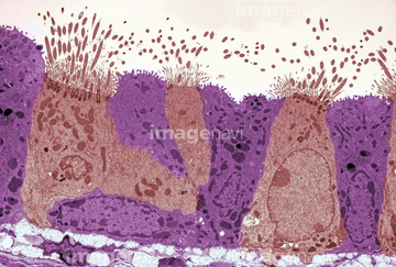
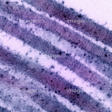
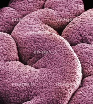
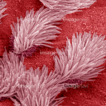
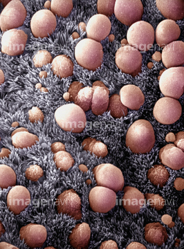
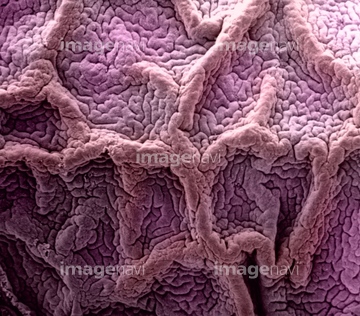
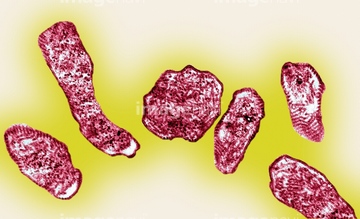
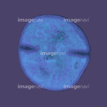
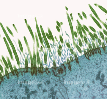
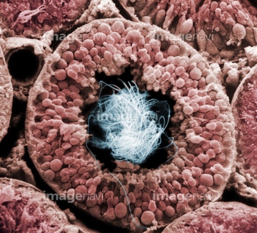
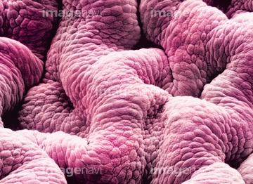
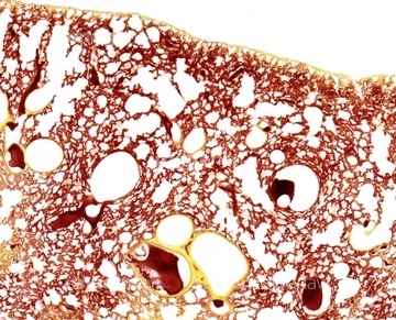
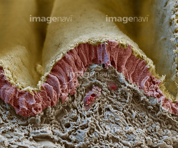
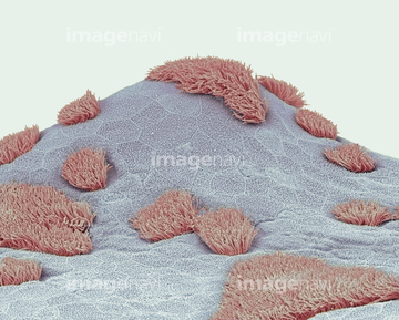
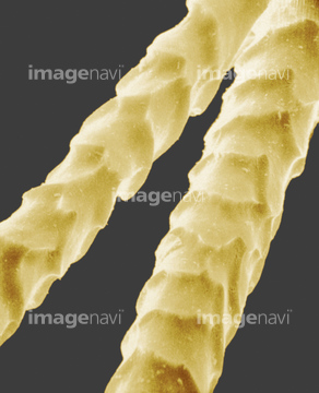
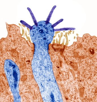
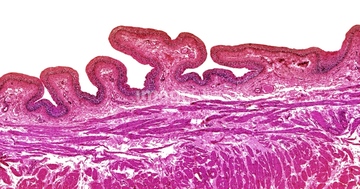
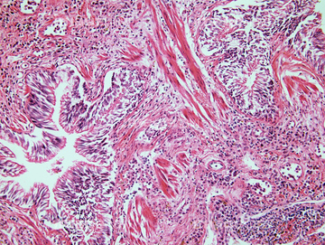
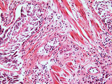
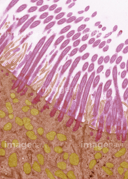
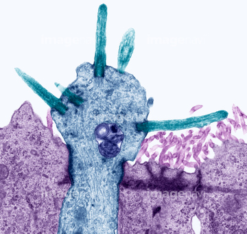
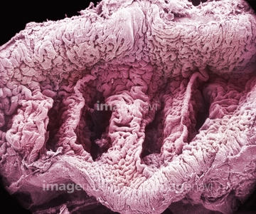
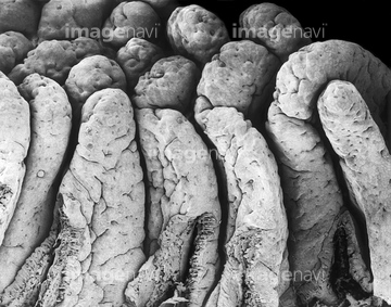
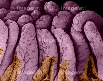
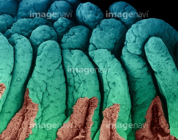
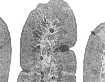
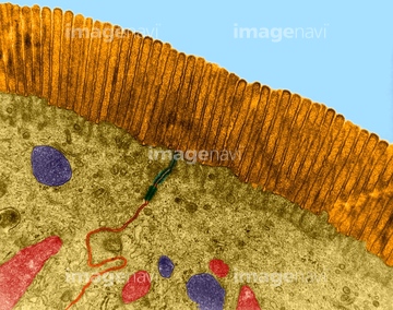
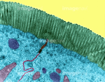
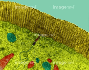
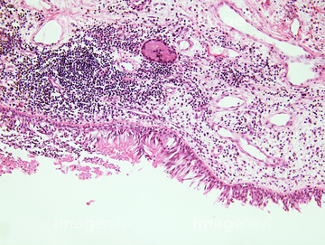
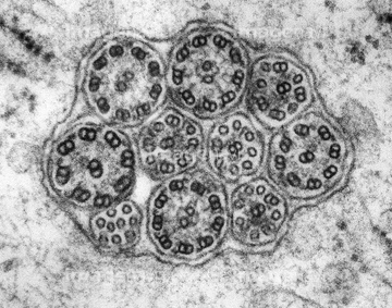
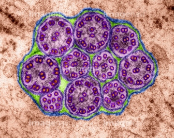
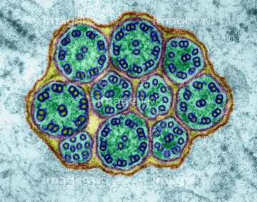
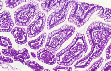
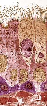
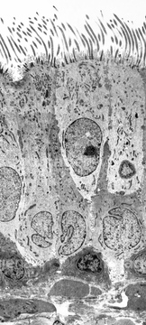
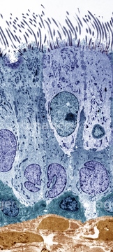
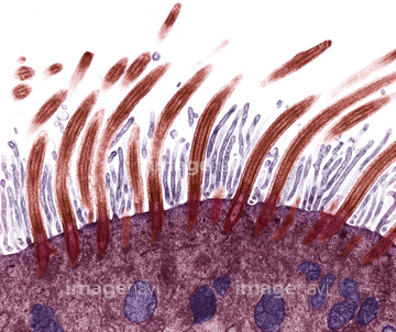
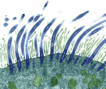
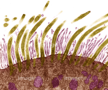
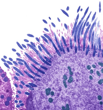
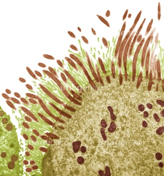
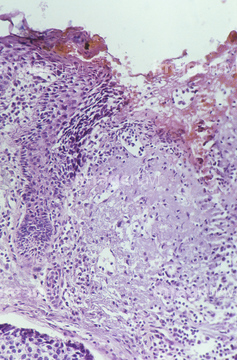
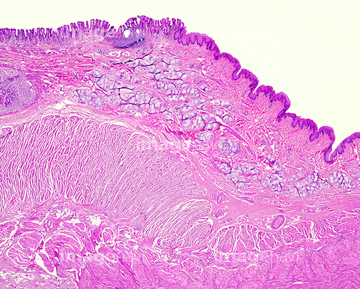
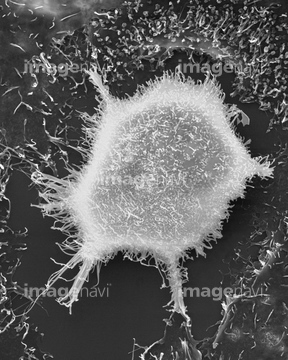
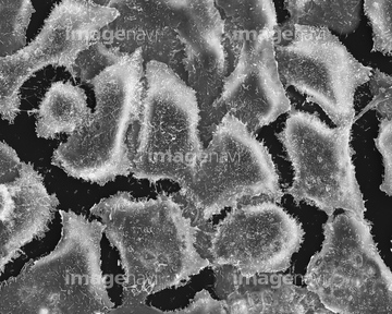
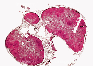
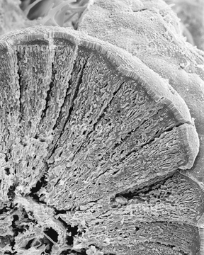
この検索結果には、Villi of the small intestine, SEM、Small intestine villus, TEM、Microvilli of the small intestine, TEM、Microvillus of the small intestine, TEM、Small intestine, microvilli, TEM、Throat cancer, light micrographなどが含まれています。
64115596
64115597
64114169
64115600
64225151
64225152

64225260
64115535
64115536

64055760
64210695
64225134
17202475
17202476
64225259
64191432
64056944
64076019
64225242
64063628
64063629
64225190
64224932
64061063
64061064
64060843
64060844
64060845
64060846
17202477
17202478
64225261
17281852
17281881
17281895
64179705
64179706
64115602
64150338
64132948
64132949
64147742
64132258

64132996
64132997
64133039
64061069
64061070
64078784
64078785
64076358

64076359

64076371
64076372

64147753
64132269
64132270
64060808
64060809
64060810
17200026
17200027
64225256
64115471
64115493
64225361
64060829
64060830
64060837
64060838
64060839
64061065
64225243

20532891
20532895
20532902
20532905
64265553
64265570
64265573
64209752
64209789
64115580
64092127
64150293
64132656
64132657
64061061
64225231
64197099
64150294
64150295
64132658
64132659
64132660
64132661
64132662
64132663
64204999
64225265
64190144
64021684
64148812

64225140
17217747
17217748
64256413
21532126
21532127
64021703

64146135

64146275
64114526
64114529
64114552
64114561
64114573
64114574
64176759
64177979
64177980
64177981
64177982
64177983
64177984
64177986
64177987
64177993
64177994
64177996
64071957
64180076
64180077
64180078
64180098
64115598
64085321
| 次ページ |
