HOME > 写真 > バックグラウンド > 色・光 > 顕微鏡写真
10,000件の写真素材が検索されました。
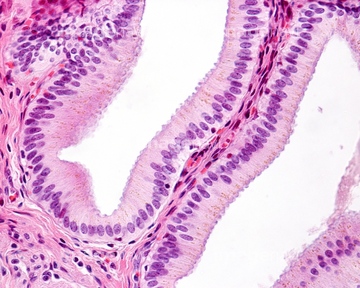
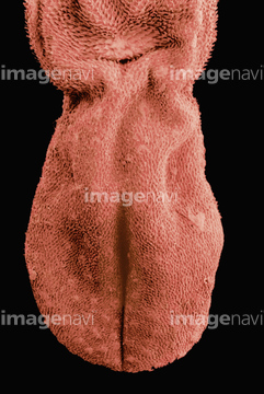
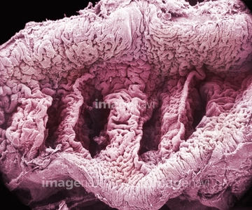
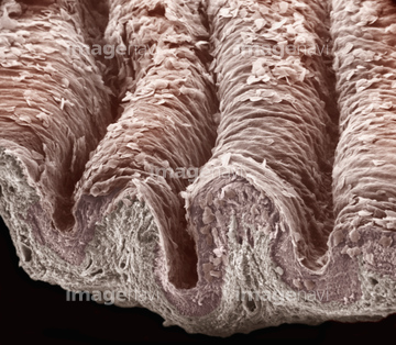
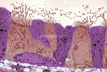
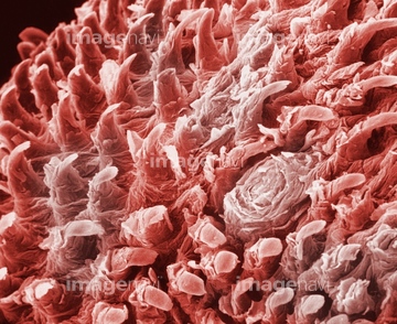
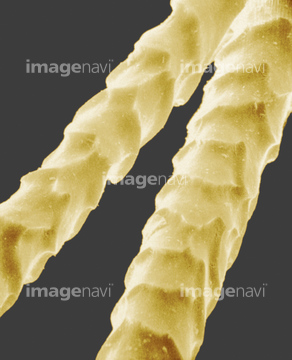
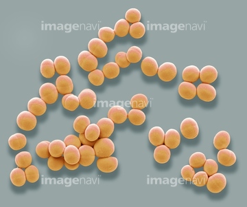
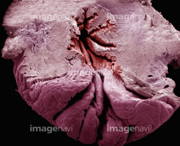
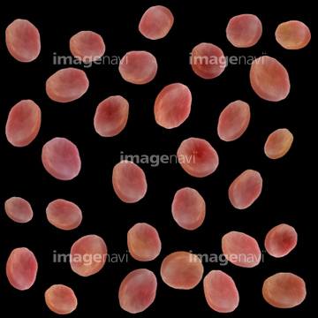
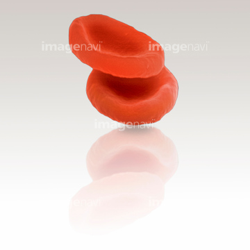
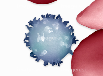
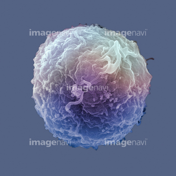
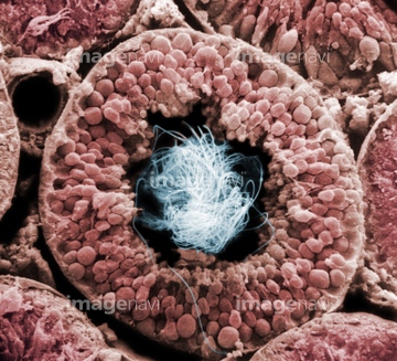
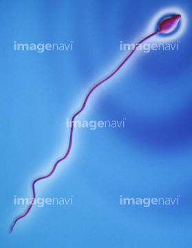
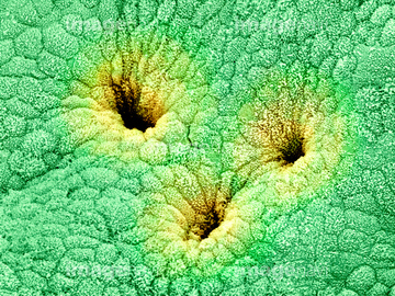
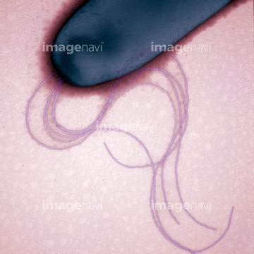
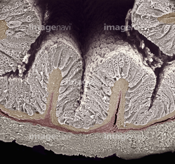
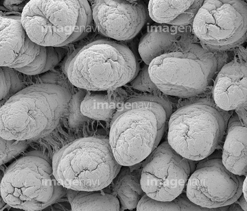
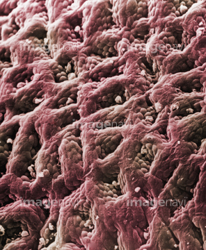
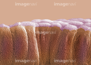
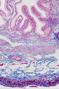
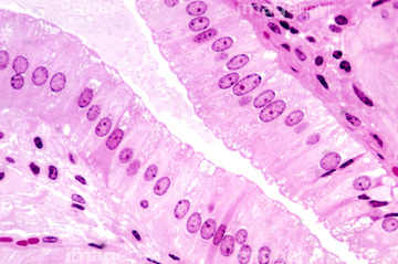
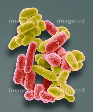
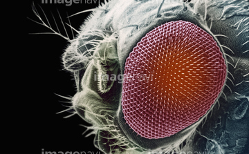
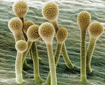
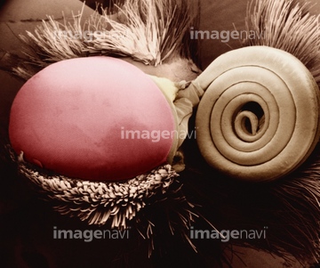
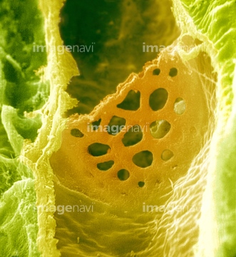
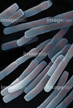
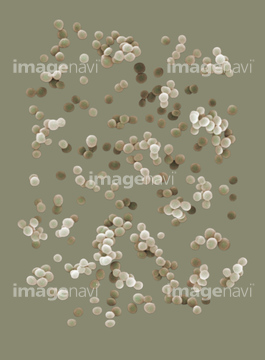
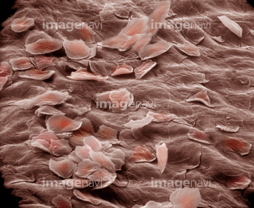
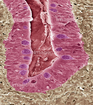
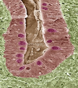
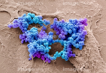
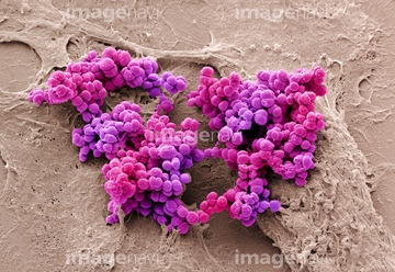
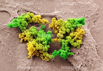
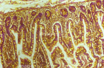
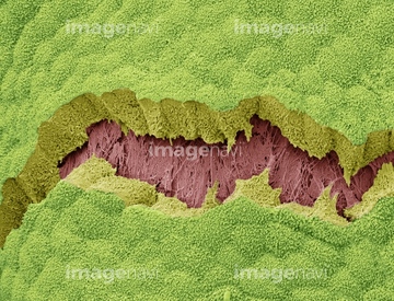
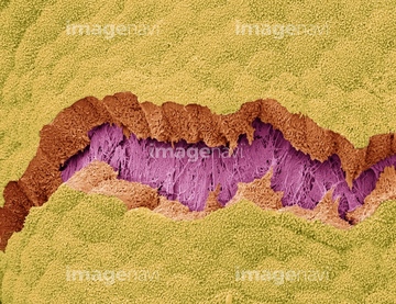
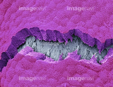
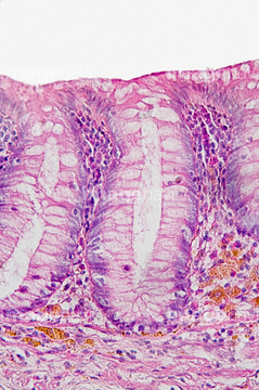
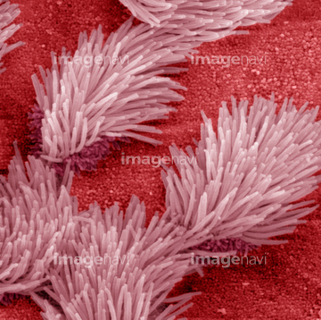
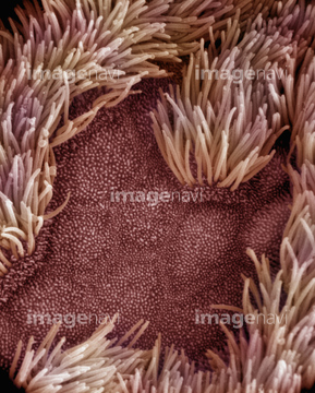
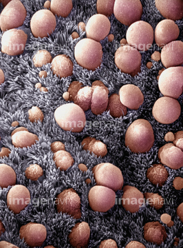
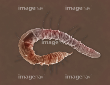
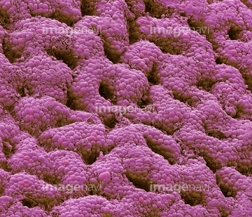
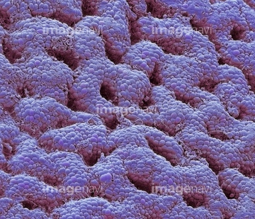
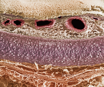
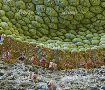
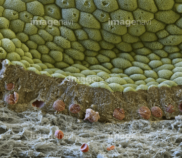
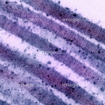
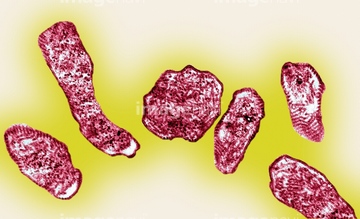
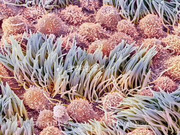
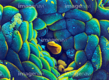
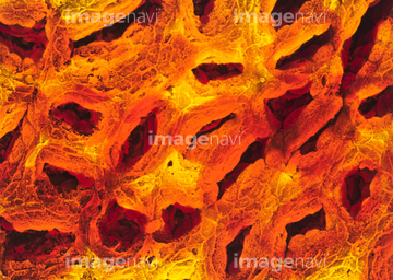
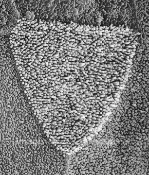
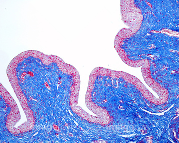
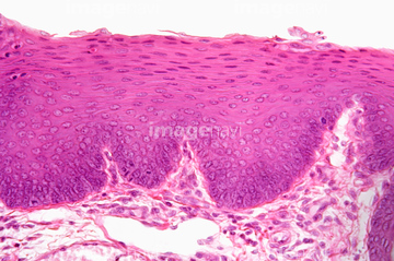
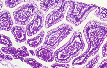
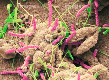
この検索結果には、Human lymphocyte white blood cell. SEM、Human testis tubule filled with sperm、Human sperm SEM X2250、Gallbladder surface, SEM、Budding Yeast SEM、Gallbladder, SEMなどが含まれています。
64225260
64225259

64146135

64146275
64221011
64225256

64225140
64225265
64225261


64225131
64225082
64115596
64225257

64225242
17281852
17281881
17281895
64234509
64234510
64115471
64115493
64115600
64225361
64076360
64076361
64224854
64123467
64123508

64225083

64225133
64115602
64152547
64225092
64225176
64234508
64224842
17246260
17246264
17246265
64115490
64115520
64115526
64115594
64115595
64225086
64225091
64225243
64225244
64225264
17200012
17200013
64136447
64225192
64114730
64224880
64225175
64225201
64008744
64008745

64104713
64104724
64104731
64104736
20528355
20528365
20528369
64197456
64114552
64114169
64225151
64225152
64196734
64197478
64206819
64235009
64235010
64066084
64071988
64071989
64114549
64114570
64114571
64115521
64225101
20523355
20523366
64225189
64143715
64143816
64115597
64056751
64088321
64088322
64150242
64114561

64055760
64123469
64123483
64123499
64236634
64236655
64225190
64115593
64143021
64143022
64143023
64008746
64008751
64055754
64114526
64114529
64114573
64114574
64143020
64143707
64143708
64143709
64143710
64143711
64143712
64143713
64143714
64143817
64143818
64143819
64143820
64143821
64161259
64161268
64115535
64115536
64225158
17201424
64084027
| 次ページ |
