HOME > 写真 > バックグラウンド > 色・光 > 顕微鏡写真
10,000件の写真素材が検索されました。
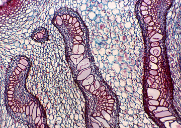
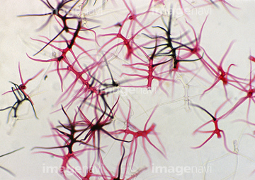
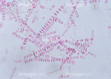
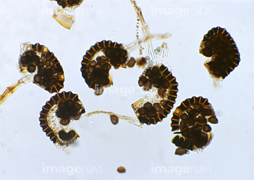
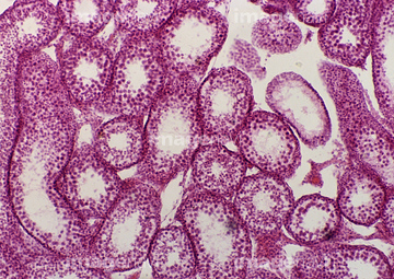
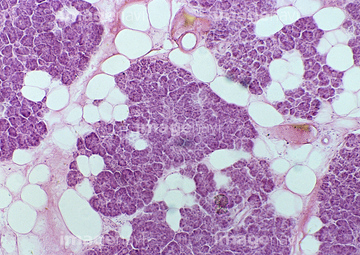
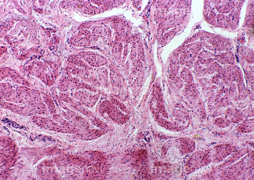
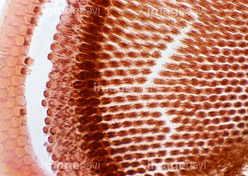
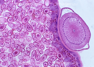
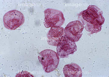
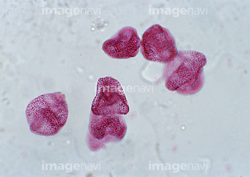
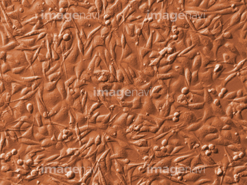
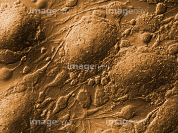
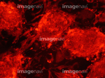
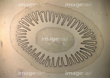
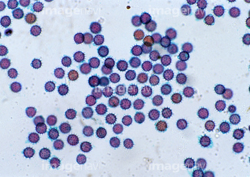
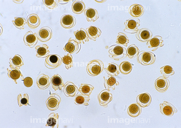
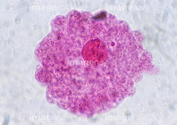
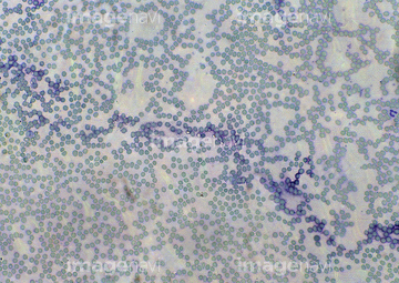
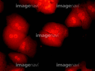
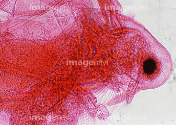
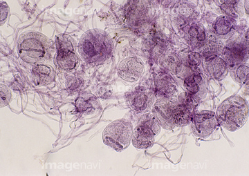
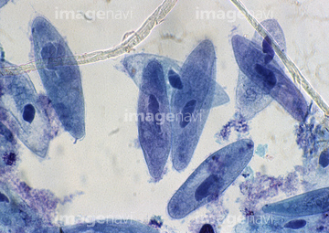
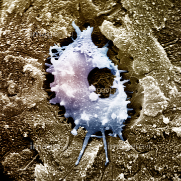
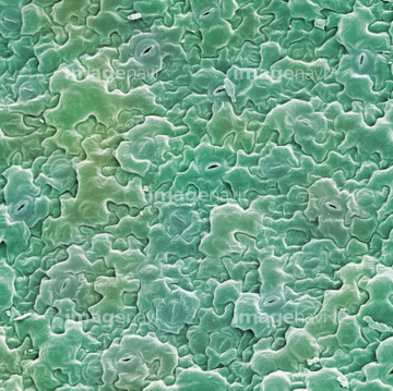
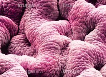
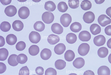
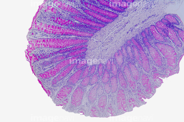
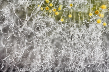
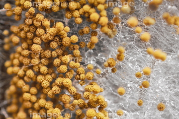
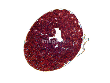
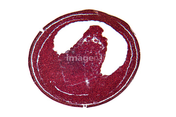
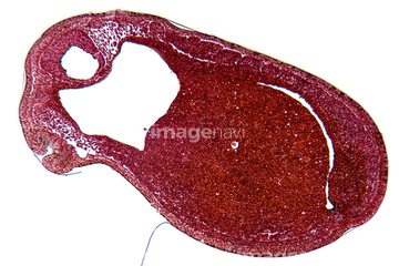
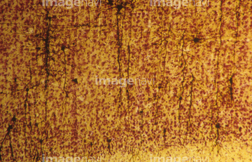
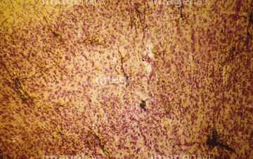
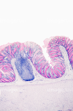
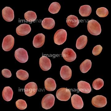
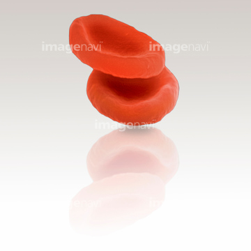
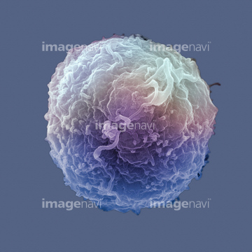
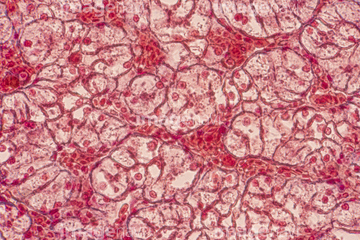
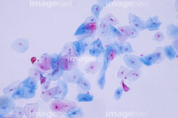
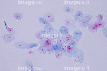
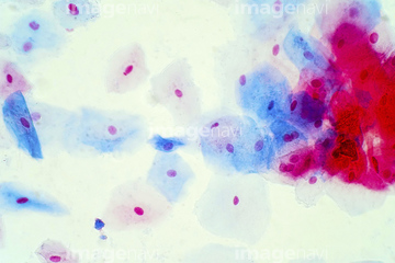
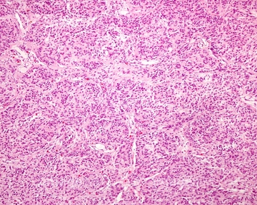
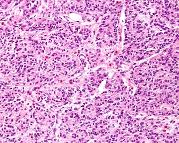
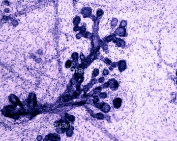
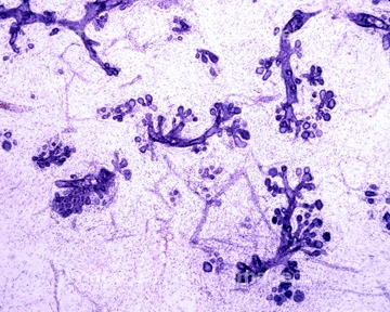
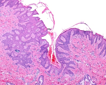
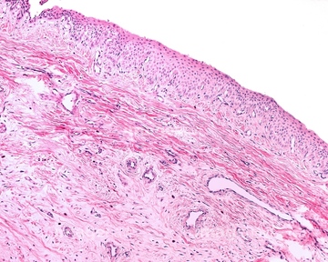
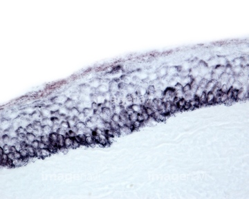
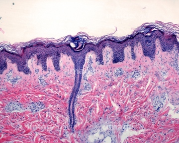
この検索結果には、細胞、肝臓の組織、赤血球、黒色腫瘍、赤色蛍光ES細胞、アメーバなどが含まれています。
99074172
99074170
99058038
00001215
00001219

99058013
99058016
99058017
99058018
99058019
99058020
99058021
99058027
99058028
99058029
99058030
99058036
99058037
99058044
99058045
99058048
99058049
99058051
99058052
99058053

99058001
99058002
99058003
99058004
99058005
99058006
99058007
99058008
99058011
99058014
99058015
99058022
99058023
99058024
99058025
99058026
99058031
99058032
99058039
99058040
99058041
99058042
99058046
99058050
99058070
00001220
99058033

99058034
00001216

10330777
99074167
99058055
99058056

99058057
99058058
99058060
99058061
99058062
99058063
99058064
99058065
99058068
99058069
10330779
10330773
10330774
00001018
99058043
99058047
99074168
99058054
10330775
10330776
99058035
99058059
99058066
99058067
99058009
99058010
99058012

70014586
99074166
99074169
99074171
10011728
00001217
00001218
10330778
64114541
64003750
64003751
64003752
64088782
64224892
64225259
64225293

17276216
17276217
17276218
17276219
17276220
17276251
17280741
17280742
64114552
64017358
64017359
64224929
64255080
64250441
64233706
64224846

64114561
64114574
64055598
64055599
64055600
64114567
64206764
64206765
64206766
64206767
64206791
64017356
64255079
64233688
64114573
64115471
64115493
64225361
64225224
20510205
20510207
20510227
64225260
64017357

64146135

64146275
64169949
64169962
64169977
64169978
| 次ページ |
